. 2011 Jun;68(11):1969-82.
doi: 10.1007/s00018-010-0544-y. Epub 2010 Oct 21.

POU homeodomain protein OCT1 modulates islet 1 expression during cardiac differentiation of P19CL6 cells

Affiliations

POU homeodomain protein OCT1 modulates islet 1 expression during cardiac differentiation of P19CL6 cells

Yinan Liu et al. Cell Mol Life Sci. 2011 Jun.

Abstract

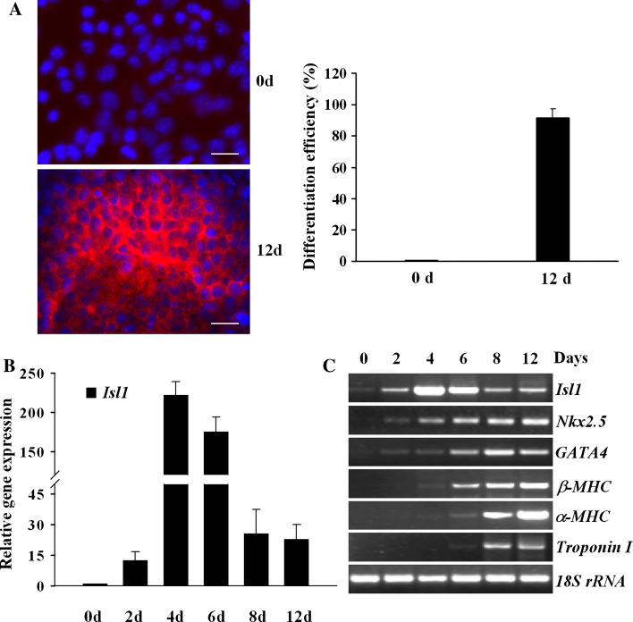
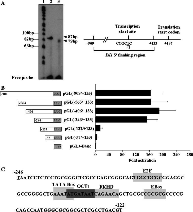
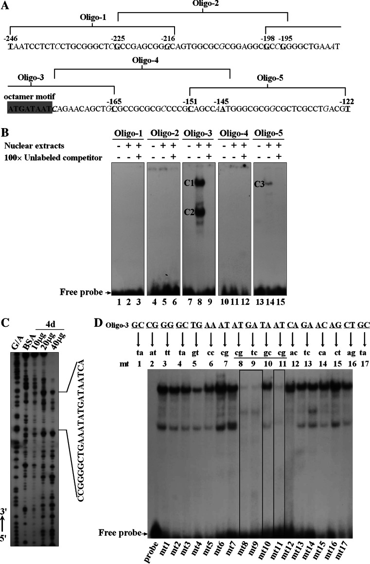
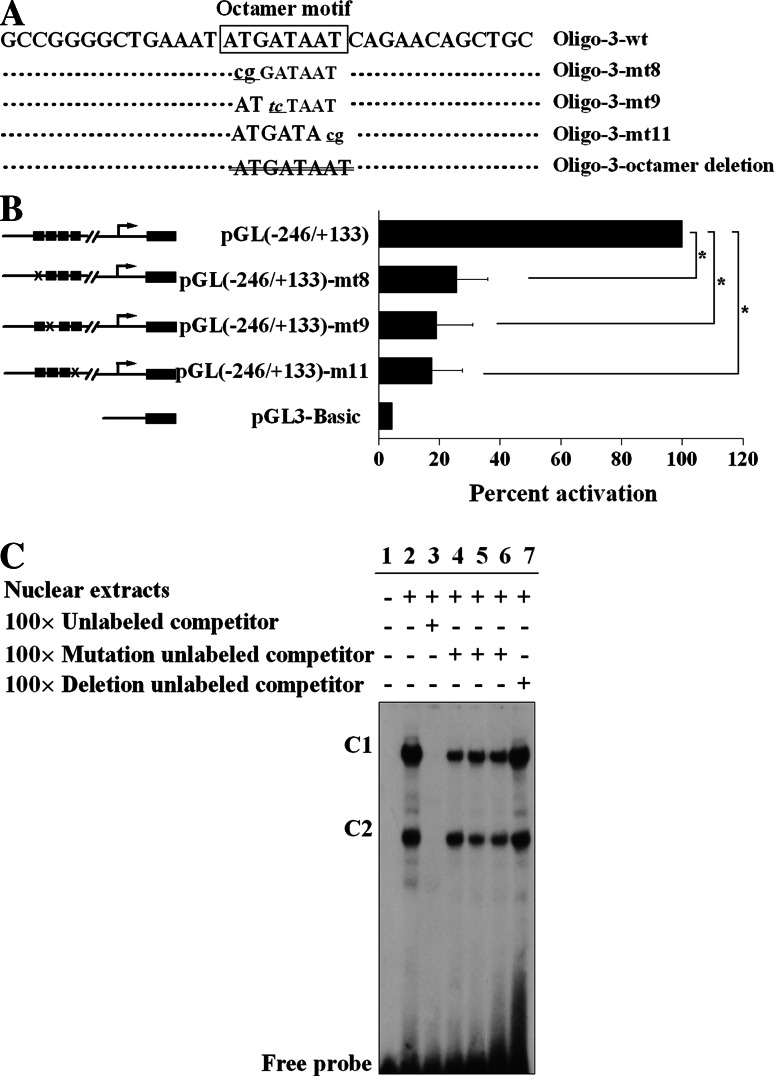
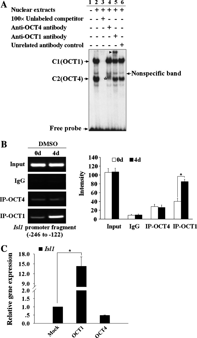
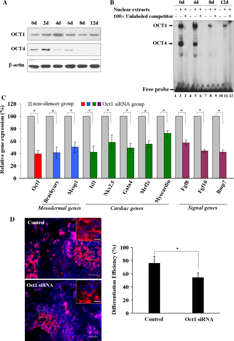
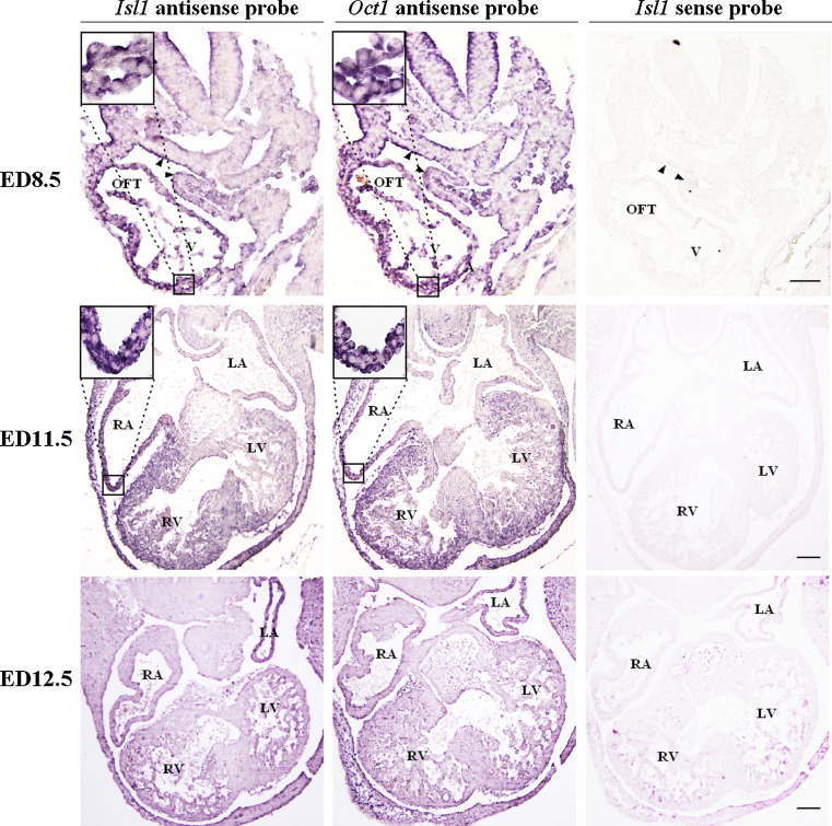
Islet 1 (ISL1), a marker of cardiac progenitors, plays a crucial role in cardiogenesis. However, the precise mechanism underlying the activation of its expression is not fully understood. Using the cardiac differentiation model of P19CL6 cells, we show that POU homeodomain protein, OCT1, modulates Isl1 expression in the process of cardiac differentiation. Oct1 knock-down resulted in reduction of Isl1 expression and downregulated mesodermal, cardiac-specific, and signal pathway gene expression. Additionally, the octamer motif located in the proximal region of Isl1 promoter is essential to Isl1 transcriptional activation. Mutation of this motif remarkably decreased Isl1 transcription. Although both OCT1 and OCT4 bound to this motif, it was OCT1 rather than OCT4 that modulated Isl1 expression. Furthermore, the correlation of OCT1 in regulation of Isl1 was revealed by in situ hybridization in early embryos. Collectively, our data highlight a novel role of OCT1 in the regulation of Isl1 expression.


Figures

Fig. 1
Fig. 1
The expression of Isl1 during cardiomyocytic differentiation of P19CL6 cells. a Uninduced and induced 12 day P19CL6 cells were analyzed by immunofluorescence staining with a monoclonal antibody against sarcomeric α-actinin (red in cytoplasm). Nuclei were counterstained with Hoechst33342 (blue). Scale bars 20 μm. Differentiation efficiency is shown on the right panel. b Real-time RT-PCR was performed to evaluate Isl1 expression during P19CL6 cell differentiation at indicated time points. Data are shown as relative Isl1 mRNA level compared with that of uninduced cells (0d) with mean ± SD from three independent experiments, each in triplicate. c RT-PCR was used to analyze the expression of Isl1 and cardiac-specific genes with RNA extracted from P19CL6 cells at indicated time points
Fig. 2
Fig. 2
Analysis of the 5′ flanking region of Isl1 gene. a Primer extension assay was used to identify the transcriptional start site of Isl1. Lane 1 ϕ×174 Hinf1 DNA marker, lane 2 control RNA, lane 3 induced day 4 P19CL6 RNA. The arrowheads indicate the products of the primer extension from control (87 bp) and experimental groups (79 bp). The map of Isl1 transcriptional start site is shown in the right panel. b Luciferase assay was performed to evaluate the activities of Isl1 promoter and its truncated constructs. Data are shown as fold activation over that of PGL3-basic promoter with means ± SD (n = 3). c Potential binding sites of transcription factors between the nucleotide −246 and −122 are indicated by boxes
Fig. 3
Fig. 3
Identification of cis elements involved in the regulation of Isl1 expression. a The 32P-ATP labeled oligonucleotides are indicated as Oligo-1, -2, -3, -4, and -5. Octamer motif is highlighted by dark gray box. b EMSA was performed using the labeled probes and nuclear extracts (NE) prepared from induced day 4 P19CL6 cells. The DNA–protein complexes formed are indicated as C1, C2, and C3. c DNase I footprinting identified the protected region by NE on the Isl1 minimal promoter. d The bases responsible for DNA binding activities on the Isl1 minimal promoter were determined by EMSA. The mutant probes are labeled as mt1 to mt17
Fig. 4
Fig. 4
The requirement of octamer motif for activation of Isl1 minimal promoter. a A series of 2 bp mutants of octamer motif (lowercase letters) and octamer deletion (crossed-out lines) from Oligo-3 were used for EMSA in c. b Luciferase assay was performed to evaluate the transcriptional activity of Isl1 minimal promoter with mutants of octamer motif. × represents two mutant bases and black boxes represent the wild-type bases. Data are shown as percent activation over that of PGL(−246/+133) with mean ± SD (n = 3) [*P < 0.05, vs. PGL(246/+133)]. c EMSA was performed to evaluate the DNA binding affinity of mutant bases within the octamer motif. The binding complexes are indicated as C1 and C2
Fig. 5
Fig. 5
Regulation of OCT1 on the Isl1 expression. a EMSA was performed using the labeled Oligo-3 with NE from induced day 4 P19CL6 cells and antibodies against OCT1 or OCT4. Addition of anti-OCT1 antibody yields a supershift band as indicted by the arrowhead (lane 5), while addition of anti-OCT4 antibody produced a weakened band as indicated by the asterisk (lane 4). Nonspecific band and free probes are also indicated by arrows. b ChIP assay was performed with antibodies against OCT1 or OCT4 in day 0 or induced day 4 P19CL6 cells. The immunoprecipitated DNA fragments were amplified by PCR for the Isl1 promoter region from −246 to +133. Input represents 10% of the total input chromatin, and IgG served as a negative control. The quantitative presentation of the bands in the left panel, based on the ChIP data in c was analyzed by Multi Gauge V3.0 software and is shown with mean ± SD (*P < 0.05) (right panel). c Real-time RT-PCR analysis of effect by overexpression of OCT1 or OCT4 on the Isl1 expression. Data are shown as relative Isl1 gene expression over that of mock with means ± SD (n = 3) (*P < 0.05, vs. mock group)
Fig. 6
Fig. 6
The influence of Oct1 knock-down on cardiogenesis during P19CL6 differentiation. a Western blot was performed to analyze OCT1 and OCT4 protein levels during P19CL6 cell differentiation at indicated time points. b EMSA was performed to assess the DNA binding activities with labeled Oligo-3 and NE from different time points of induced P19CL6 cells. c Real-time RT-PCR was used to evaluate a series of mesodermal (blue), cardiac-specific (green), and signal pathway genes (purple) expression in P19CL6 cells under the condition of Oct1 knock-down (as shown in red bar) at induced day 4. Data are normalized to 18S expression and presented as percent expression versus that of the nonsilencer group with mean ± SD (n = 3) (*P < 0.05). d Immunofluorescence staining with a monoclonal antibody against sarcomeric α-actinin (red) was analyzed after Oct1 knock-down. Nuclei were counterstained with Hoechst33342 (blue). The outlined areas are shown magnified in the upper right corners. Scale bars 10 μm. Differentiation efficiency is expressed as the percentage of α-actinin-positive cells out of the total number of cells counted (right panel) (*P < 0.05)
Fig. 7
Fig. 7
In situ hybridization analysis of Isl1 and Oct1 mRNAs on the embryos at ED8.5, ED11.5, and ED12.5. Serial sections of embryos were hybridized in situ with an antisense probe of Isl1 or Oct1. Strong positive signals were seen in SM, OFT, and V at ED8.5 and RA, RV, and a specific area of LV at ED11.5, but no signals were detected at ED12.5. Corresponding sense probes were used as negative controls. The results of Isl1 sense probe are shown. Black triangles indicate SM. The outlined areas are shown in the upper left corners. Scale bars 100 μm. SM Splanchnic mesoderm, OFT outflow tract, A atria, LA left atria, RA right atria, V ventricles, RV right ventricle, LV left ventricle

References

    1. Karlsson O, Thor S, Norberg T, Ohlsson H, Edlund T. Insulin gene enhancer binding protein Isl-1 is a member of a novel class of proteins containing both a homeo- and a Cys-His domain. Nature. 1990;344:879–882. doi: 10.1038/344879a0. - DOI - PubMed
    1. Pfaff SL, Mendelsohn M, Stewart CL, Edlund T, Jessell TM. Requirement for LIM homeobox gene Isl1 in motor neuron generation reveals a motor neuron-dependent step in interneuron differentiation. Cell. 1996;84:309–320. doi: 10.1016/S0092-8674(00)80985-X. - DOI - PubMed
    1. Ahlgren U, Pfaff SL, Jessell TM, Edlund T, Edlund H. Independent requirement for ISL1 in formation of pancreatic mesenchyme and islet cells. Nature. 1997;385:257–260. doi: 10.1038/385257a0. - DOI - PubMed
    1. Du A, Hunter CS, Murray J, Noble D, Cai CL, Evans SM, Stein R, May CL. Islet 1 is required for the maturation, proliferation, and survival of the endocrine pancreas. Diabetes. 2009;58:2059–2069. doi: 10.2337/db08-0987. - DOI - PMC - PubMed
    1. Cai CL, Liang X, Shi Y, Chu PH, Pfaff SL, Chen J, Evans S. Isl1 identifies a cardiac progenitor population that proliferates prior to differentiation and contributes a majority of cells to the heart. Dev Cell. 2003;5:877–889. doi: 10.1016/S1534-5807(03)00363-0. - DOI - PMC - PubMed

Publication types

MeSH terms

Substances

LinkOut - more resources