. 2010 Nov;105(6):771-85.
doi: 10.1007/s00395-010-0124-1. Epub 2010 Oct 20.

Inhibition of permeability transition pore opening by mitochondrial STAT3 and its role in myocardial ischemia/reperfusion

Affiliations

Inhibition of permeability transition pore opening by mitochondrial STAT3 and its role in myocardial ischemia/reperfusion

Kerstin Boengler et al. Basic Res Cardiol. 2010 Nov.

Abstract

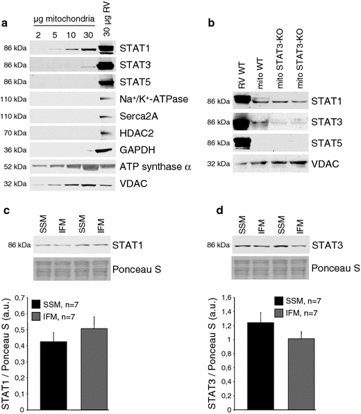
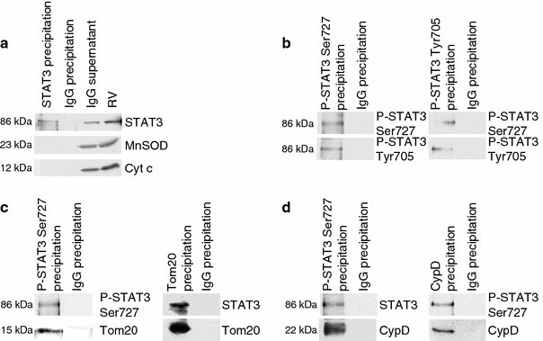
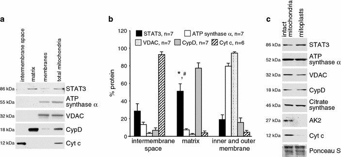
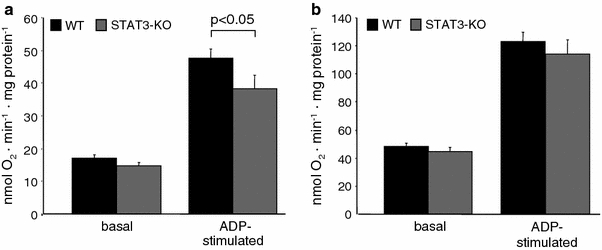
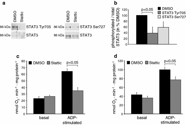
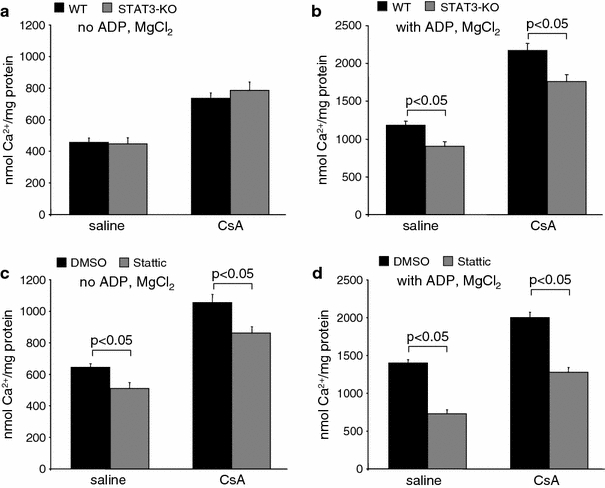
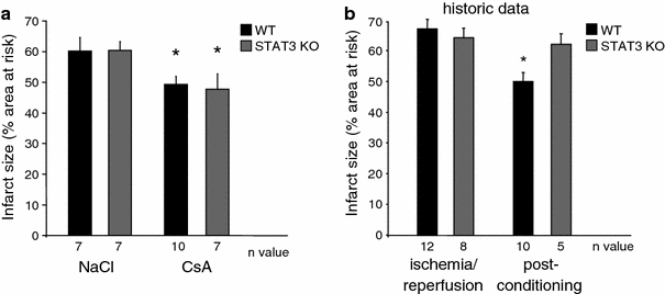
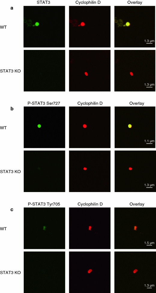
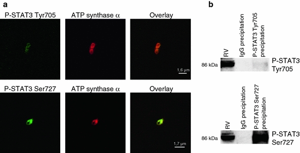
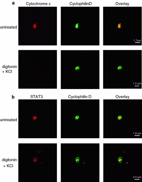
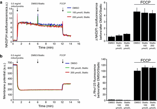
The signal transducer and activator of transcription 3 (STAT3) contributes to cardioprotection by ischemic pre- and postconditioning. Mitochondria are central elements of cardioprotective signaling, most likely by delaying mitochondrial permeability transition pore (MPTP) opening, and STAT3 has recently been identified in mitochondria. We now characterized the mitochondrial localization of STAT3 and its impact on respiration and MPTP opening. STAT3 was mainly present in the matrix of subsarcolemmal and interfibrillar cardiomyocyte mitochondria. STAT1, but not STAT5 was also detected in mitochondria under physiological conditions. ADP-stimulated respiration was reduced in mitochondria from mice with a cardiomyocyte-specific deletion of STAT3 (STAT3-KO) versus wildtypes and in rat mitochondria treated with the STAT3 inhibitor Stattic (STAT3 inhibitory compound, 6-Nitrobenzo[b]thiophene 1,1-dioxide). Mitochondria from STAT3-KO mice and Stattic-treated rat mitochondria tolerated less calcium until MPTP opening occurred. STAT3 co-immunoprecipitated with cyclophilin D, the target of the cardioprotective agent and MPTP inhibitor cyclosporine A (CsA). However, CsA reduced infarct size to a similar extent in wildtype and STAT3-KO mice in vivo. Thus, STAT3 possibly contributes to cardioprotection by stimulation of respiration and inhibition of MPTP opening.


Figures

Fig. 1
Fig. 1
STAT1 and 3 are present in LV mitochondria. a Western blot analysis was performed for STAT1, 3, and 5 and marker proteins of subcellular compartments (Na+/K+-ATPase: sarcolemma, Serca2a: sarcoplasmic reticulum, HDAC2: nucleus, GAPDH: cytosol, ATP synthase α: mitochondria, VDAC: mitochondria) on rat total right ventricular (RV) and different amounts of LV SSM protein extracts (n = 4). Western blots were exposed to X-ray films such that similar immunoreactivities were obtained for STAT1, 3, and 5 in RV protein extracts. b Western blot analysis was performed for STAT1, 3, 5, and the mitochondrial marker protein VDAC on 25 μg total right ventricular (RV) and LV SSM proteins from WT and STAT3-KO mice (n = 4). c Western blot analysis was performed for STAT1 on SSM and IFM isolated from rat myocardium. Ponceau S staining is shown to demonstrate equal protein loading. The amount of STAT1 was normalized to Ponceau S staining of myocardial SSM and IFM. d Western blot analysis was performed for STAT3 on SSM and IFM isolated from rat myocardium. Ponceau S staining is shown to demonstrate equal protein loading. The amount of STAT3 was normalized to Ponceau S staining of myocardial SSM and IFM
Fig. 2
Fig. 2
Identification of mitochondrial proteins which co-immunoprecipitate with STAT3 and characterization of the dual mitochondrial STAT3 phosphorylation. Total STAT3 was immunoprecipitated from rat LV SSM. The specificity of the immunoprecipitation is indicated by the lack of STAT3 in the IgG precipitation as well as by the lack of MnSOD and cytochrome c (Cyt C) immunoreactivities (a). Phosphorylated STAT3 [Ser727 and Tyr705 (b)], as well as Tom20 (c) or cyclophilin D (CypD, d) were immunoprecipitated from rat SSM proteins. Rabbit IgGs were used as negative control. Western blot analysis was performed for total or phosphorylated STAT3 (Ser727 and Tyr705), Tom20, and CypD (n = 3–6)
Fig. 3
Fig. 3
STAT3 is present in the mitochondrial matrix. a Rat LV SSM were subfractionated into the proteins of the intermembrane space, the matrix, and the inner and outer membrane (membranes). Western blot analysis was performed for STAT3 and marker proteins of different submitochondrial compartments (intermembrane space: Cyt c, matrix: CypD, outer membrane: VDAC, inner membrane: ATP synthase α). Total mitochondrial protein extracts were used as control. bBar graphs representing the percentage of protein in mitochondrial compartments. The total amount of pixels from each subfractionation protocol was set as 100%. *,#p < 0.05 for STAT3 immunoreactivity in the matrix versus the intermembrane space and the inner and outer membrane, respectively. c Rat LV SSM were incubated in hypo-osmotic buffer to induce mitochondrial swelling and subsequently characterized by western blot analysis. Swelling induces loss of marker proteins for the intermembrane space (adenylate kinase 2, AK2, and cytochrome C, Cyt c), whereas immunoreactivities for membrane proteins (ATP synthase α, inner membrane) or VDAC (outer membrane), matrix proteins [citrate synthase, cyclophilin D (CypD)] as well as STAT3 immunoreactivity were unchanged
Fig. 4
Fig. 4
Genetic reduction of STAT3 decreases mitochondrial ADP-stimulated oxygen consumption. Oxygen consumption of WT (n = 12) and STAT3-KO (n = 12) SSM was measured under basal conditions and after addition of 40 μmol/L ADP using glutamate/malate as substrates for complex 1 (a) or succinate with rotenone as substrate for complex 2 (b)
Fig. 5
Fig. 5
Pharmacological inhibition of STAT3 decreases mitochondrial ADP-stimulated oxygen consumption. a Total STAT3 was immunoprecipitated from purified rat LV SSM which were incubated with 100 μmol/L Stattic or DMSO. Western blot analysis was performed for STAT3 phosphorylated at tyrosine 705, serine 727 or total STAT3 (n = 4). The ratio of phosphorylated over total STAT3 in Stattic-treated mitochondria is shown in percentage of DMSO-treated control mitochondria (b). Oxygen consumption was measured under basal conditions and after addition of 40 μM ADP to rat LV SSM incubated for 1 h at 4°C with DMSO or 100 μmol/L Stattic using glutamate/malate as substrates for complex 1 (c, n = 6) or succinate with rotenone as substrate for complex 2 (d, n = 6)
Fig. 6
Fig. 6
Genetic reduction and pharmacological inhibition of STAT3 enhance calcium-induced MPTP opening. The calcium concentration at which opening of the MPTP occurred (increase in calcium green 5N fluorescence) was calculated in STAT3-KO or WT mouse SSM in the presence or absence of CsA using a substrates for complex 1 without ADP and MgCl2 (WT: n = 7, STAT3-KO: n = 9) or b with ADP and MgCl2 (WT: n = 10, STAT3-KO: n = 11), and in Stattic- or DMSO-treated rat SSM in the presence or absence of CsA using c substrates for complex 1 without ADP and MgCl2 (n = 9) or d with ADP and MgCl2 (n = 7)
Fig. 7
Fig. 7
STAT3 is important for infarct size reduction by ischemic postconditioning, but not for pharmacological postconditioning with cyclosporine A (CsA). a Infarct size (in % of the area at risk) was determined in WT and STAT3-KO mice undergoing 30 min ischemia and 120 min reperfusion. 10 mg/kg CsA or 0.9% NaCl solution were administered intravenously 5 min before reperfusion, *p < 0.05. b Historic data [2] showing that ischemic postconditioning by 3 cycles of 10 s ischemia and reperfusion each at the onset of reperfusion reduced infarct size after 30 min ischemia and 120 min reperfusion in WT, but not in STAT3-KO mice, *p < 0.05
Fig. 8
Fig. 8
Mitochondrial STAT3 content is reduced in aged mouse hearts. Western blot analysis was performed for STAT3 on LV SSM from young (8 weeks) and aged (21 months) wildtype mice. As positive control served the reduced mitochondrial content of connexin 43 and as negative control the unchanged protein content of ATP synthase α
Fig. 9
Fig. 9
Total and phosphorylated STAT3 are present in mouse left ventricular (LV) mitochondria. a LV subsarcolemmal mitochondria (SSM) of WT (n = 6) and STAT3-KO mice (n = 8) were stained with antibodies against STAT3 (green) and the mitochondrial marker cyclophilin D (red) and were analyzed by confocal laser scan microscopy. Overlay pixels are shown in yellow. Whereas in SSM isolated from the LV of WT mice about 91% (347 mitochondria counted) of the ATP synthase positive mitochondria were also positive for STAT3, only 13% (248 mitochondria counted) of the SSM had immunoreactivity for STAT3 in STAT3-KO mice. b SSM from the LV of WT (n = 4) and STAT3-KO mice (n = 4) were stained with antibodies against STAT3 phosphorylated at Ser727 (green) and the mitochondrial marker cyclophilin D (red) and were analyzed by confocal laser scan microscopy. Overlay pixels are shown in yellow. c SSM from the LV of WT (n = 4) and STAT3-KO mice (n = 4) were stained with antibodies against STAT3 phosphorylated at Tyr705 (green) and the mitochondrial marker cyclophilin D (red) and were analyzed by confocal laser scan microscopy. Overlay pixels are shown in yellow
Fig. 10
Fig. 10
Mitochondrial STAT3 is phosphorylated. a Subsarcolemmal mitochondria (SSM) from the rat LV were stained with antibodies against STAT3 phosphorylated at Tyr705 or Ser727 (green) and the mitochondrial marker ATP synthase α (red) and were analyzed by confocal laser scan microscopy (n = 4). Overlay pixels are shown in yellow. b STAT3 phosphorylated at Tyr705 or Ser727 was immunoprecipitated from rat SSM proteins. IgGs were used as negative control. Western blot analysis on immunoprecipitated proteins was performed for serine or tyrosine phosphorylated STAT3 (n = 4). To compare signal intensities for STAT3 phosphorylated at Tyr705 or Ser727, western blots were exposed to X-ray films such that the signal intensities for phosphorylated STAT3 in total right ventricular (RV) protein extracts were similar
Fig. 11
Fig. 11
STAT3 is present in the matrix of left ventricular (LV) subsarcolemmal mitochondria (SSM). a LV rat SSM (untreated or treated with digitonin and KCl in order to remove proteins from the outer membrane and the intermembrane space) were stained with antibodies against cytochrome c (intermembrane space, red) and cyclophilin D (matrix, green), and were analyzed by confocal laser scan microscopy. Overlay pixels are shown in yellow (n = 3). b Untreated or digitonin/KCl-treated SSM were stained with antibodies against STAT3 (red) and cyclophilin D (green) and were analyzed by confocal laser scan microscopy. Overlay pixels are shown in yellow (n = 3)
Fig. 12
Fig. 12
Stattic causes no acute toxic effects on mitochondria. a NAD(P)H autofluorescence of 0.5 mg/mL rat left ventricular (LV) subsarcolemmal mitochondria (SSM) was measured before and after the addition of 100 or 200 μmol/L Stattic or DMSO as vehicle. To induce collapse of the mitochondrial membrane potential, 100 nmol/L FCCP was given. The difference between the NAD(P)H autofluorescence 1 min before and 1 min after the addition of Stattic, DMSO or FCCP, respectively, was calculated (n = 5). b Mitochondrial membrane potential of 0.5 mg/mL rat LV SSM was measured with rhodamine 123 before and after the addition of 100 or 200 μmol/L Stattic or DMSO as vehicle. The difference between the fluorescence 1 min before and 1 min after the addition of Stattic, DMSO or FCCP, respectively, was calculated (n = 5)

References

    1. Argaud L, Gateau-Roesch O, Raisky O, Loufouat J, Robert D, Ovize M. Postconditioning inhibits mitochondrial permeability transition. Circulation. 2005;111:194–197. doi: 10.1161/01.CIR.0000151290.04952.3B. - DOI - PubMed
    1. Boengler K, Buechert A, Heinen Y, Roeskes C, Hilfiker-Kleiner D, Heusch G, Schulz R. Cardioprotection by ischemic postconditioning is lost in aged and STAT3-deficient mice. Circ Res. 2008;102:131–135. doi: 10.1161/CIRCRESAHA.107.164699. - DOI - PubMed
    1. Boengler K, Dodoni G, Rodriguez-Sinovas A, Cabestrero A, Ruiz-Maena M, Gres P, Konietzka I, Lopez-Iglesias C, García-Dorado D, Heusch G, Schulz R. Connexin 43 in cardiomyocyte mitochondria and its increase by ischemic preconditioning. Cardiovasc Res. 2005;67:234–244. doi: 10.1016/j.cardiores.2005.04.014. - DOI - PubMed
    1. Boengler K, Hilfiker-Kleiner D, Drexler H, Heusch G, Schulz R. The myocardial JAK/STAT pathway: from protection to failure. Pharmacol Therap. 2008;120:172–185. doi: 10.1016/j.pharmthera.2008.08.002. - DOI - PubMed
    1. Boengler K, Konietzka I, Buechert A, Heinen Y, Garcia-Dorado D, Heusch G, Schulz R. Loss of ischemic preconditioning’s cardioprotection in aged mouse hearts is associated with reduced gap junctional and mitochondrial levels of connexin 43. Am J Physiol Heart Circ Physiol. 2007;292:H1764–H1769. doi: 10.1152/ajpheart.01071.2006. - DOI - PubMed

Publication types

MeSH terms