

Age specific responses to acute inhalation of diffusion flame soot particles: cellular injury and the airway antioxidant response
- PMID: 20961279
- PMCID: PMC3111018
- DOI: 10.3109/08958378.2010.513403
Age specific responses to acute inhalation of diffusion flame soot particles: cellular injury and the airway antioxidant response
Abstract
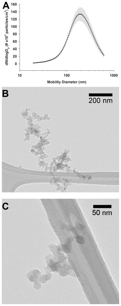
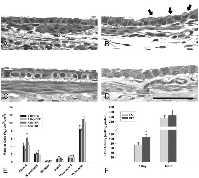
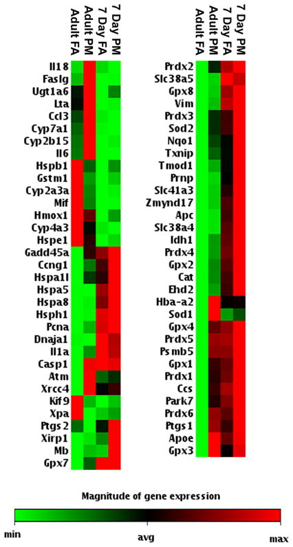
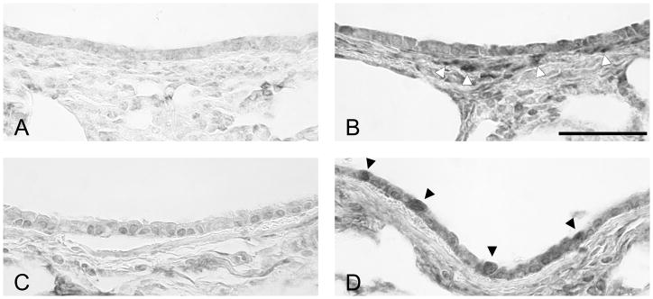
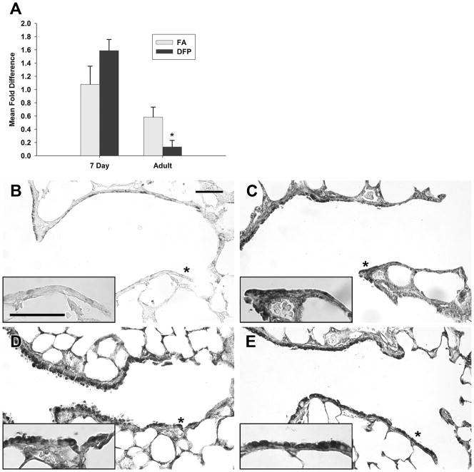
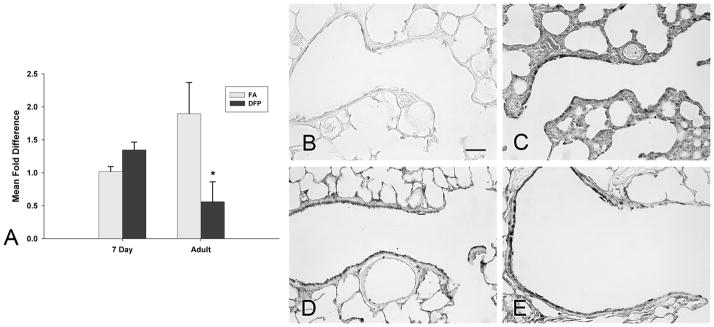
Current studies of particulate matter (PM) are confounded by the fact that PM is a complex mixture of primary (crustal material, soot, metals) and secondary (nitrates, sulfates, and organics formed in the atmosphere) compounds with considerable variance in composition by sources and location. We have developed a laboratory-based PM that is replicable, does not contain dust or metals and that can be used to study specific health effects of PM composition in animal models. We exposed both neonatal (7 days of age) and adult rats to a single 6-h exposure of laboratory generated fine diffusion flame particles (DFP; 170 µg/m(3)), or filtered air. Pulmonary gene and protein expression as well as indicators of cytotoxicity were evaluated 24 h after exposure. Although DFP exposure did not alter airway epithelial cell composition in either neonates or adults, increased lactate dehydrogenase activity was found in the bronchoalveolar lavage fluid of neonates indicating an age-specific increase in susceptibility. In adults, 16 genes were differentially expressed as a result of DFP exposure whereas only 6 genes were altered in the airways of neonates. Glutamate cysteine ligase protein was increased in abundance in both DFP exposed neonates and adults indicating an initiation of antioxidant responses involving the synthesis of glutathione. DFP significantly decreased catalase gene expression in adult airways, although catalase protein expression was increased by DFP in both neonates and adults. We conclude that key airway antioxidant enzymes undergo changes in expression in response to a moderate PM exposure that does not cause frank epithelial injury and that neonates have a different response pattern than adults.
Figures

Similar articles
-
Susceptibility to inhaled flame-generated ultrafine soot in neonatal and adult rat lungs.Toxicol Sci. 2011 Dec;124(2):472-86. doi: 10.1093/toxsci/kfr233. Epub 2011 Sep 13. Toxicol Sci. 2011. PMID: 21914721 Free PMC article.
-
Oxidative injury in the lungs of neonatal rats following short-term exposure to ultrafine iron and soot particles.J Toxicol Environ Health A. 2010;73(12):837-47. doi: 10.1080/15287391003689366. J Toxicol Environ Health A. 2010. PMID: 20391124
-
Age-specific effects on rat lung glutathione and antioxidant enzymes after inhaling ultrafine soot.Am J Respir Cell Mol Biol. 2013 Jan;48(1):114-24. doi: 10.1165/rcmb.2012-0108OC. Epub 2012 Oct 11. Am J Respir Cell Mol Biol. 2013. PMID: 23065132 Free PMC article.
-
Mechanisms of particulate matter toxicity in neonatal and young adult rat lungs.Res Rep Health Eff Inst. 2008 Oct;(135):3-41; discussion 43-52. Res Rep Health Eff Inst. 2008. PMID: 19203021
-
Combustion-derived flame generated ultrafine soot generates reactive oxygen species and activates Nrf2 antioxidants differently in neonatal and adult rat lungs.Part Fibre Toxicol. 2013 Aug 1;10:34. doi: 10.1186/1743-8977-10-34. Part Fibre Toxicol. 2013. PMID: 23902943 Free PMC article.
Cited by
-
Outdoor Air Pollution and New-Onset Airway Disease. An Official American Thoracic Society Workshop Report.Ann Am Thorac Soc. 2020 Apr;17(4):387-398. doi: 10.1513/AnnalsATS.202001-046ST. Ann Am Thorac Soc. 2020. PMID: 32233861 Free PMC article.
-
Susceptibility to inhaled flame-generated ultrafine soot in neonatal and adult rat lungs.Toxicol Sci. 2011 Dec;124(2):472-86. doi: 10.1093/toxsci/kfr233. Epub 2011 Sep 13. Toxicol Sci. 2011. PMID: 21914721 Free PMC article.
-
Emulating Near-Roadway Exposure to Traffic-Related Air Pollution via Real-Time Emissions from a Major Freeway Tunnel System.Environ Sci Technol. 2022 Mar 2:10.1021/acs.est.1c07047. doi: 10.1021/acs.est.1c07047. Online ahead of print. Environ Sci Technol. 2022. PMID: 35235290 Free PMC article.
-
Combustion derived ultrafine particles induce cytochrome P-450 expression in specific lung compartments in the developing neonatal and adult rat.Am J Physiol Lung Cell Mol Physiol. 2013 May 15;304(10):L665-77. doi: 10.1152/ajplung.00370.2012. Epub 2013 Mar 15. Am J Physiol Lung Cell Mol Physiol. 2013. PMID: 23502512 Free PMC article.
-
Onset of alveolar recirculation in the developing lungs and its consequence on nanoparticle deposition in the pulmonary acinus.J Appl Physiol (1985). 2016 Jan 1;120(1):38-54. doi: 10.1152/japplphysiol.01161.2014. Epub 2015 Oct 22. J Appl Physiol (1985). 2016. PMID: 26494453 Free PMC article.
References
-
- ALA. American Lung Association State of the Air: 2009. American Lung Association; New York, NY: 2009.
-
- Baker GL, Shultz MA, Fanucchi MV, Morin DM, Buckpitt AR, Plopper CG. Assessing gene expression in lung subcompartments utilizing in situ RNA preservation. Toxicol Sci. 2004;77:135–141. - PubMed
-
- Billet S, Garcon G, Dagher Z, Verdin A, Ledoux F, Cazier F, Courcot D, Aboukais A, Shirali P. Ambient particulate matter (PM2.5): physicochemical characterization and metabolic activation of the organic fraction in human lung epithelial cells (A549) Environ Res. 2007;105:212–223. - PubMed
-
- Branis M, Safranek J, Hytychova A. Exposure of children to airborne particulate matter of different size fractions during indoor physical education at school. Building and Environment. 2009;44:1246–1252.
Publication types
MeSH terms
Substances
Grants and funding
LinkOut - more resources
Full Text Sources
Medical