. 2011 Jun;117(3):110-22.
doi: 10.1016/j.bandl.2010.09.008. Epub 2010 Oct 18.

Support for anterior temporal involvement in semantic error production in aphasia: new evidence from VLSM

Affiliations

Support for anterior temporal involvement in semantic error production in aphasia: new evidence from VLSM

Grant M Walker et al. Brain Lang. 2011 Jun.

Abstract

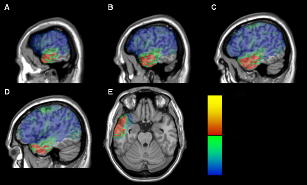
Semantic errors in aphasia (e.g., naming a horse as "dog") frequently arise from faulty mapping of concepts onto lexical items. A recent study by our group used voxel-based lesion-symptom mapping (VLSM) methods with 64 patients with chronic aphasia to identify voxels that carry an association with semantic errors. The strongest associations were found in the left anterior temporal lobe (L-ATL), in the mid- to anterior MTG region. The absence of findings in Wernicke's area was surprising, as were indications that ATL voxels made an essential contribution to the post-semantic stage of lexical access. In this follow-up study, we sought to validate these results by re-defining semantic errors in a manner that was less theory dependent and more consistent with prior lesion studies. As this change also increased the robustness of the dependent variable, it made it possible to perform additional statistical analyses that further refined the interpretation. The results strengthen the evidence for a causal relationship between ATL damage and lexically-based semantic errors in naming and lend confidence to the conclusion that chronic lesions in Wernicke's area are not causally implicated in semantic error production.


Figures

Fig. 1
Fig. 1
Maps depicting lesion overlaps of the 64 subjects in the left hemisphere. Maps A–D are at MNI x coordinates of −64, −58, −52, and −46 respectively. Map E is a single axial slice at z=−24. The regions of maximal overlap (bright yellow) are in the peri-Sylvian regions, particularly anterior. Only voxels lesioned in at least five subjects were included, yielding 404, 565 total voxels to be tested.
Fig. 2
Fig. 2
Maps of the reliability (t statistic) of the difference in SemPlus between patients with and without lesions in each voxel (rendered on the MNI-space ch2 volume). Voxels exceeding the FDR threshold (q=0.01) are rendered in a red (t=3.09) to yellow (t>6.18) scale, while nonsignificant values are rendered on a scale from green (t just below threshold) to blue (t=0 or below). Maps A–D are at MNI x coordinates of −64, −58, −52, and −46 respectively. Map E is a single axial slice at z=−24. The peak value of 8.79 was held in common by four contiguous voxels centered at MNI coordinates −64.5, −3.75, −24.5 – the anterior cortex of the middle temporal gyrus (Maps A and E show crosshairs at −64, −4, −24). There were 9 (of 64) subjects who had lesions encroaching the peak voxels.
Fig. 3
Fig. 3
Maps of the reliability (t statistic) of the difference in SemPlus, after factoring out PhonErr, between patients with and without lesions in each voxel (rendered on the MNI-space ch2 volume). Voxels exceeding the FDR threshold (q=0.01) are rendered in a red (t=3.08) to yellow (t>6.16) scale, while non-significant values are rendered on a scale from green (t just below threshold) to blue (t=0 or below). Maps A–D are at MNI x coordinates of −64, −58, −52, and −46 respectively. Map E is a single axial slice at z=−24. The centroid of the peak voxels (t=8.54) in the map were at −64.5, −3.75, −24.5 (exactly the same region as in the unfiltered analysis).
Fig. 4
Fig. 4
Maps of the reliability (t statistic) of the difference in SemPlus, after factoring out NVcomp, between patients with and without lesions in each voxel (rendered on the MNI-space ch2 volume). Voxels exceeding the FDR threshold (q=0.01) are rendered in a red (t=3.18) to yellow (t>6.35) scale, while non-significant values are rendered on a scale from green (t just below threshold) to blue (t=0 or below). Maps A–D are at MNI x coordinates of −64, −58, −52, and −46 respectively. Map E is a single axial slice at z=−24. The centroid of the peak voxels (t=7.25) in the map were at −64.5, −3.75, −24.5 (exactly the same region as in the unfiltered analysis).
Fig. 5
Fig. 5
Maps of the reliability (t statistic) of the difference in SemPlus, after factoring out total lesion volume, between patients with and without lesions in each voxel (rendered on the MNI-space ch2 volume). Voxels exceeding the FDR threshold (q=0.01) are rendered in a red (t=3.54) to yellow (t>7.09) scale, while non-significant values are rendered on a scale from green (t just below threshold) to blue (t=0 or below). Maps A–D are at MNI x coordinates of −64, −58, −52, and −46 respectively. Map E is a single axial slice at z=−24. The peak statistical value of 5.95 is held by four contiguous voxels at MNI coordinates −62.5, −3, −27.75 (a very slight shift from the unfiltered analysis).
Fig. 6
Fig. 6
Maps of the reliability (t statistic) of the difference in SemPlus, after factoring out percent damage to BA22, between patients with and without lesions in each voxel (rendered on the MNI-space ch2 volume). Voxels exceeding the FDR threshold (q=0.01) are rendered in a red (t=3.53) to yellow (t>7.06) scale, while non-significant values are rendered on a scale from green (t just below threshold) to blue (t=0 or below). Maps A–D are at MNI x coordinates of −64, −58, −52, and −46 respectively. Map E is a single axial slice at z=−24. The peak statistical value of 5.86 is held by four contiguous voxels at MNI coordinates −62.5, −3, −27.75 (the same as the total lesion volume filtered analysis).

References

    1. Antonucci SM, Beeson PM, Labiner DM, Rapcsak SZ. Lexical retrieval and semantic knowledge in patients with left inferior temporal lobe lesions. Aphasiology. 2008;22(3):281–304. - PMC - PubMed
    1. Antonucci SM, Beeson PM, Rapcsak SZ. Anomia in patients with left inferior temporal lobe lesions. Aphasiology. 2004;18(5/6/7):543–554. - PMC - PubMed
    1. Avants B, Schoenemann PT, Gee JC. Lagrangian frame diffeomorphic image registration: Morphometric comparison of human and chimpanzee cortex. Medical Image Analysis. 2006;10:397–412. - PubMed
    1. Bedny M, McGill M, Thompson-Schill SL. Semantic adaptation and competition during word comprehension. Cerebral Cortex. 2008;18(11):2574–2585. - PMC - PubMed
    1. Benson DF, Ardila A. Aphasia: A clinical perspective. New York, NY: Oxford University Press; 1996.