. 2011 Apr 6;8(57):500-5.
doi: 10.1098/rsif.2010.0413. Epub 2010 Oct 20.

Nano-mechanical properties of individual mineralized collagen fibrils from bone tissue

Affiliations

Nano-mechanical properties of individual mineralized collagen fibrils from bone tissue

Fei Hang et al. J R Soc Interface. .

Abstract

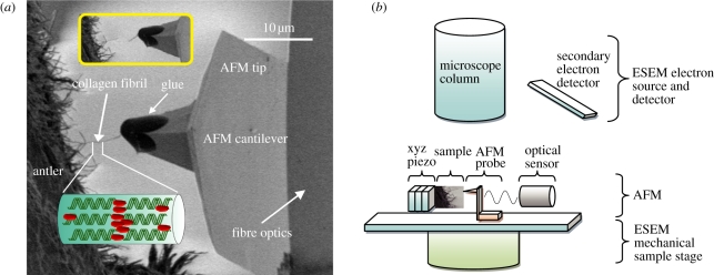
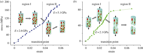
Mineralized collagen fibrils (MCFs) are distinct building blocks for bone material and perform an important mechanical function. A novel experimental technique using combined atomic force microscopy and scanning electron microscopy is used to manipulate and measure the mechanical properties of individual MCFs from antler, which is a representative bone tissue. The recorded stress-strain response of individual MCFs under tension shows an initial linear deformation region for all fibrils, followed by inhomogeneous deformation above a critical strain. This inhomogeneous deformation is indicative of fibrils exhibiting either yield or strain hardening and suggests possible mineral compositional changes within each fibril. A phenomenological model is used to describe the fibril nano-mechanical behaviour.


Figures

Figure 1.
Figure 1.
(a) SEM image showing a typical testing configuration. A large number of exposed CFs observed at the fracture surface of antler bone. An individual CF protruding from the fracture surface is attached to the glue at the end of the AFM probe. Translation of the AFM probe away from the fibril causes tensile deformation of the fibril until failure occurs, which is shown in the inset image. (b) Schematic showing set-up of combined SEM–AFM. (Online version in colour.)
Figure 2.
Figure 2.
Water content in samples varies with different exposure time to the vacuum conditions of the SEM instrument. The water content was measured as weight lost during the TGA tests when heating the samples up to 200°C.
Figure 3.
Figure 3.
(a) The stress–strain plot individual MCFs. All MCFs show a linear stress–strain behaviour with a modulus of 2.4 ± 0.4 GPa in region I, before the transition point. Beyond the transition in region II, MCFs exhibit inhomogeneous stress–strain deformation behaviour as indicated by the shaded area. Inhomogeneous deformation of MCFs is through either a lowering of the modulus indicating yield or an enhancement of the modulus. (b) Failure strength versus ultimate strain to failure of MCFs in tension. The arrow indicates ‘brittle-like’ behaviour of increasing MCF strength with decreasing failure strain. (Online version in colour.)
Figure 4.
Figure 4.
SEM back-scattered image of the antler bone fracture surface. The light regions on the image indicate higher mineralization. The corresponding Ca/P ratio in the image varied from 1.48 to 3.12, indicating hydroxyapatite mineral is present [22,23], and the content varies throughout the antler bone.
Figure 5.
Figure 5.
Tensile stress–strain curves for MCFs showing two distinct mechanical behaviours. Tropocollagen uncoiling occurs initially in both types of fibrils within region I. In (a), the fibril shows an enhanced elastic modulus in region II owing to mineral increasing the stress transfer between tropocollagen macromolecules. In (b), the low mineral density within the fibrils allows sliding between tropocollagen molecules, resulting in a plastic deformation. (Online version in colour.)

References

    1. Currey J. D. 1999. The design of mineralised hard tissues for their mechanical functions. J. Exp. Biol. 202, 3285–3294 - PubMed
    1. Rho J. Y., Kuhn-Spearing L., Zioupos P. 1998. Mechanical properties and the hierarchical structure of bone. Med. Eng. Phys. 20, 92–10210.1016/S1350-4533(98)00007-1 (doi:10.1016/S1350-4533(98)00007-1) - DOI - DOI - PubMed
    1. Weiner S., Traub W., Wagner H. D. 1999. Lamellar bone: structure–function relations. J. Struct. Biol. 126, 241–25510.1006/jsbi.1999.4107 (doi:10.1006/jsbi.1999.4107) - DOI - DOI - PubMed
    1. Rajaram A., Ramanathan N. 1982. Tensile properties of antler bone. Calcif. Tissue Int. 34, 301–30510.1007/BF02411255 (doi:10.1007/BF02411255) - DOI - DOI - PubMed
    1. Zioupos P., Currey J. D., Sedman A. J. 1994. An examination of the micromechanics of failure of bone and antler by acoustic emission tests and laser scanning confocal microscopy. Med. Eng. Phys. 16, 203–21210.1016/1350-4533(94)90039-6 (doi:10.1016/1350-4533(94)90039-6) - DOI - DOI - PubMed

LinkOut - more resources