. 2011 Feb;336(2):313-20.
doi: 10.1124/jpet.110.174904. Epub 2010 Oct 20.

The nitric oxide prodrug JS-K is effective against non-small-cell lung cancer cells in vitro and in vivo: involvement of reactive oxygen species

Affiliations

The nitric oxide prodrug JS-K is effective against non-small-cell lung cancer cells in vitro and in vivo: involvement of reactive oxygen species

Anna E Maciag et al. J Pharmacol Exp Ther. 2011 Feb.

Abstract

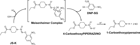
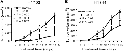
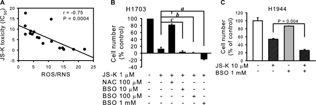
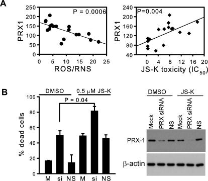
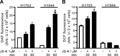
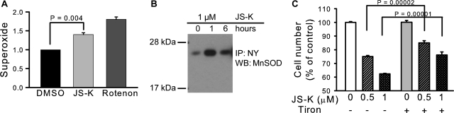
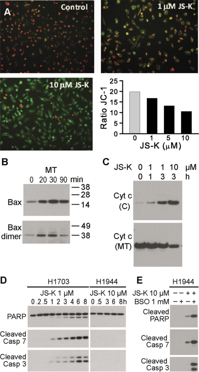
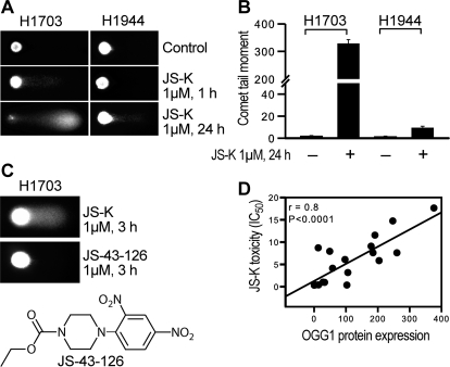
Non-small-cell lung cancer is among the most common and deadly forms of human malignancies. Early detection is unusual, and there are no curative therapies in most cases. Diazeniumdiolate-based nitric oxide (NO)-releasing prodrugs are a growing class of promising NO-based therapeutics. Here, we show that O(2)-(2,4-dinitrophenyl)-1-[(4-ethoxycarbonyl)piperazin-1-yl]diazen-1-ium-1,2-diolate (JS-K) is a potent cytotoxic agent against a subset of human non-small-cell lung cancer cell lines both in vitro and as xenografts in mice. JS-K treatment led to 75% reduction in the growth of H1703 lung adenocarcinoma cells in vivo. Differences in sensitivity to JS-K in different lung cancer cell lines seem to be related to their endogenous levels of reactive oxygen species (ROS)/reactive nitrogen species (RNS). Other related factors, levels of peroxiredoxin 1 (PRX1) and 8-oxo-deoxyguanosine glycosylase (OGG1), also correlated with drug sensitivity. Treatment of the lung adenocarcinoma cells with JS-K resulted in oxidative/nitrosative stress in cells with high basal levels of ROS/RNS, which, combined with the arylating properties of the compound, was reflected in glutathione depletion and alteration in cellular redox potential, mitochondrial membrane permeabilization, and cytochrome c release. Inactivation of manganese superoxide dismutase by nitration was associated with increased superoxide and significant DNA damage. Apoptosis followed these events. Taken together, the data suggest that diazeniumdiolate-based NO-releasing prodrugs may have application as a personalized therapy for lung cancers characterized by high levels of ROS/RNS. PRX1 and OGG1 proteins, which can be easily measured, could function as biomarkers for identifying tumors sensitive to the therapy.


Figures

Fig. 1.
Fig. 1.
Structure of JS-K and scheme of nitric oxide release upon reaction with GSH. DNP-SG, S-(2,4-dinitrophenyl)glutathione.
Fig. 2.
Fig. 2.
JS-K significantly reduced growth of NSCLC cells in vivo. JS-K was administered intravenously at 6 μmol/kg, three times a week for 3 weeks, and tumors were measured with a caliper. Growth of both cell lines, JS-K-sensitive H1703 (A) and over 10-fold less sensitive H1944 (B), was inhibited. Values are medians, and the relevant 95% confidence interval bars are shown (Mann-Whitney test). Letters indicate the significance of the differences between JS-K-treated and control mice at each time point. The treatment did not affect body weights. The average body weight for all mice was 22.7 ± 0.25 g (mean ± S.E.) at the beginning of the experiment. At the termination, the average weights of the control groups were 24.57 ± 0.88 g (n = 11) and 24.41 ± 0.57 g (n = 13), for H1703 and H1944 xenograft studies, respectively. The weights of JS-K treated animals were 24.92 ± 0.39 g (n = 9) and 24.42 ± 0.56 g (n = 11), for the H1703 and H1944 xenograft groups, respectively.
Fig. 3.
Fig. 3.
JS-K toxicity depends on intracellular ROS/RNS. A, JS-K toxicity (as IC50 values) correlated significantly with endogenous ROS/RNS levels, measured as DCF fluorescence (arbitrary units). B, pretreatment with NAC reduced JS-K toxicity, whereas depletion of glutathione using the glutathione synthase inhibitor BSO sensitized the cells to JS-K. The H1703 cells were treated with NAC or BSO for 24 h, followed by 24 h with 1 μM JS-K. The number of surviving cells was assessed by the MTT assay. a, P < 0.0001; b, P = 0.003; c, P = 0.04 by Mann-Whitney test. C, depletion of GSH in the H1944 cells sensitized the cells to JS-K. Cells were treated with 1 mM BSO in complete medium for 24 h followed by 10 μM JS-K for another 24 h, and the number of surviving cells was assessed by the MTT assay.
Fig. 4.
Fig. 4.
A, protein levels of PRX1 (expressed relative to the HPL1D cell line used as an internal control) correlated negatively with intracellular ROS/RNS level measured as DCF fluorescence (arbitrary units). JS-K toxicity measured as IC50 values correlated with levels of PRX1. B, silencing of peroxiredoxins with a pool of siRNAs sensitized the cells to JS-K. H1703 cells were treated with a siRNA pool for all six isoforms of PRX for 48 h in complete medium followed by 0.5 μM JS-K for 24 h. Percentage of dead cells was measured by the trypan blue exclusion method. Loss of PRX-1 signal after siRNA treatment was shown by Western blot. M, mock control; si, siRNA to PRX pool; NS, nonsilencing siRNA negative control.
Fig. 5.
Fig. 5.
A, intracellular NO release after JS-K treatment measured as DAF fluorescence. B, intracellular ROS/RNS level after JS-K treatment measured as DCF fluorescence. A representative experiment is shown (n = 4). a, P < 0.01; b, P < 0.001; c, P < 0.0001, by paired t test, compared with cells treated with DMSO only.
Fig. 6.
Fig. 6.
JS-K-induced increase in mitochondrial superoxide and MnSOD tyrosine nitration. A, mitochondrial superoxide level measured with MitoSOX fluorescence. Rotenone was used as positive control. B, H1703 cells were treated with 1 μM JS-K for 1 or 6 h. The level of 3-nitrotyrosine (NY) in MnSOD was assessed by immunoprecipitation (IP) with an anti-nitrotyrosine antibody followed by immunoblot (WB) with an antibody to MnSOD. C, the superoxide scavenger Tiron had a protective effect against JS-K toxicity; H1703 cells were pretreated with 10 mM Tiron in complete medium for 1 h, followed by 24 h with JS-K and then the cell number was assessed by MTT assay.
Fig. 7.
Fig. 7.
A, mitochondrial membrane potential (ΔΨm) was analyzed using JC-1 mitochondrial membrane dye. H1703 cells were treated with DMSO (control) or JS-K (1–10 μM) for 1 h. The drug caused an increase in the green (JC-1 monomers) and decrease in the red fluorescence (JC-1 aggregates) indicative of loss of ΔΨm. Ratio of JC-1 (red to green) was calculated. B, immunoblot demonstrating increase in total Bax and Bax dimers in mitochondrial fraction (MT) within 20 min after 1 μM JS-K treatment. C, cytochrome c increase in the cytosol (C) and decrease in the mitochondria (MT) of H1703 cells after JS-K treatment. D, PARP cleavage and effector caspase 3 and 7 activation as shown by Western blot, in the H1703 cells, but not in JS-K-resistant H1944 cells, even at a 10-fold higher concentration of the drug. E, pretreatment with pro-oxidant BSO for 16 h sensitized H1944 cells for apoptosis after 24 h treatment with JS-K.
Fig. 8.
Fig. 8.
DNA strand break damage analyzed by comet assay. A, comparison of comet signals in H1703 and H1944 cells after JS-K treatment. B, comet tail moment was quantified using CometScore software (TriTek Inc., Sumerduck, VA). C, JS-43-126, a non-NO-releasing JS-K analog (seen in the scheme), did not cause DNA strand break damage. D, OGG1 levels correlated significantly with JS-K toxicity measured as IC50 value.

References

    1. Bagci EZ, Vodovotz Y, Billiar TR, Ermentrout B, Bahar I. (2008) Computational insights on the competing effects of nitric oxide in regulating apoptosis. PloS One 3:e2249. - PMC - PubMed
    1. Bentz BG, Hammer ND, Milash B, Klein S, Burnett DM, Radosevich JA, Haines GK., 3rd (2007) The kinetics and redox state of nitric oxide determine the biological consequences in lung adenocarcinoma. Tumour Biol 28:301–311 - PubMed
    1. Bentz BG, Hammer ND, Radosevich JA, Haines GK., 3rd (2004) Nitrosative stress induces DNA strand breaks but not caspase mediated apoptosis in a lung cancer cell line. J Carcinog 3:16. - PMC - PubMed
    1. Bonavida B, Baritaki S, Huerta-Yepez S, Vega MI, Chatterjee D, Yeung K. (2008) Novel therapeutic applications of nitric oxide donors in cancer: roles in chemo- and immunosensitization to apoptosis and inhibition of metastases. Nitric Oxide 19:152–157 - PubMed
    1. Bonavida B, Khineche S, Huerta-Yepez S, Garbán H. (2006) Therapeutic potential of nitric oxide in cancer. Drug Resist Updat 9:157–173 - PubMed

Publication types

MeSH terms

Substances