. 2010 Dec;24(12):2253-66.
doi: 10.1210/me.2009-0448. Epub 2010 Oct 20.

Cytokine and progesterone receptor interplay in the regulation of MUC1 gene expression

Affiliations

Cytokine and progesterone receptor interplay in the regulation of MUC1 gene expression

Neeraja Dharmaraj et al. Mol Endocrinol. 2010 Dec.

Abstract

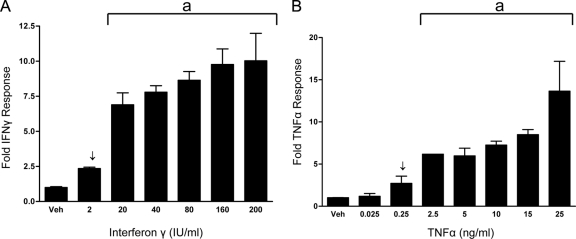
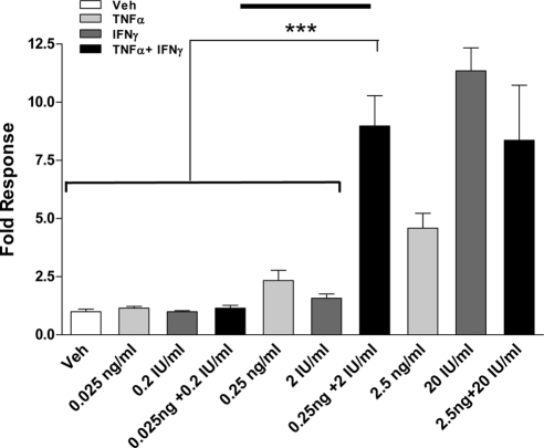
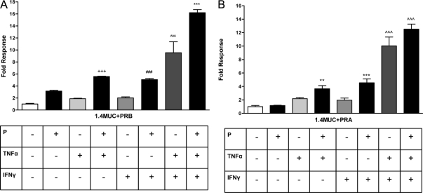
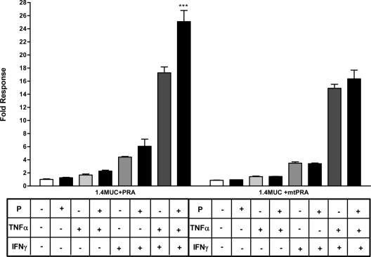
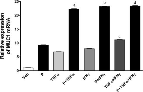
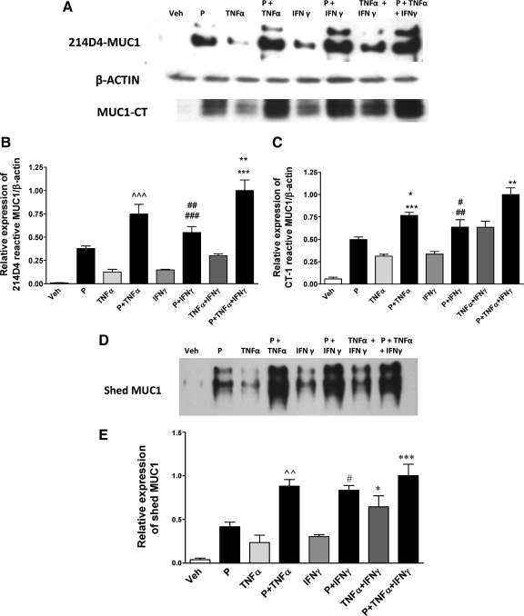
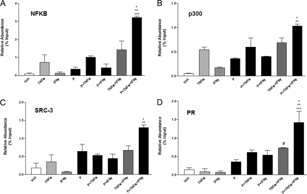
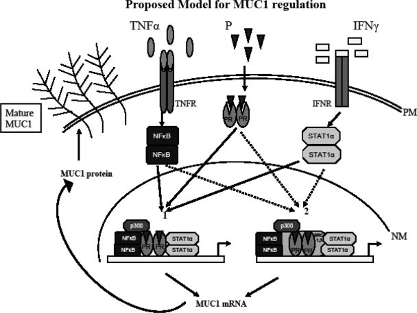
Mucin 1 (MUC1), a transmembrane mucin expressed at the apical surface of uterine epithelia, is a barrier to microbial infection and enzymatic attack. MUC1 loss at implantation sites appears to be required to permit embryo attachment and implantation in most species. MUC1 expression is regulated by progesterone (P) and proinflammatory cytokines, including TNFα and interferon γ (IFNγ). TNFα and IFNγ are highly expressed in uterine tissues under conditions where MUC1 expression is also high and activate MUC1 expression via their downstream transcription factors, nuclear factor (NF) κB and signal transducers and activators of transcription. P receptor (PR) regulates MUC1 gene expression in a PR isoform-specific fashion. Here we demonstrate that interactions among PR isoforms and cytokine-activated transcription factors cooperatively regulate MUC1 expression in a human uterine epithelial cell line, HES. Low doses of IFNγ and TNFα synergistically stimulate MUC1 promoter activity, enhance PRB stimulation of MUC1 promoter activity and cooperate with PRA to stimulate MUC1 promoter activity. Cooperative stimulation of MUC1 promoter activity requires the DNA-binding domain of the PR isoforms. MUC1 mRNA and protein expression is increased by cytokine and P treatment in HES cells stably expressing PRB. Using chromatin immunoprecipitation assays, we demonstrate efficient recruitment of NFκB, p300, SRC3 (steroid receptor coactivator 3), and PR to the MUC1 promoter. Collectively, our studies indicate a dynamic interplay among cytokine-activated transcription factors, PR isoforms and transcriptional coregulators in modulating MUC1 expression. This interplay may have important consequences in both normal and pathological contexts, e.g. implantation failure and recurrent miscarriages.


Figures

Figure 1
Figure 1
MUC1 promoter responsiveness to IFNγ (A) or TNFα (B) in HES cells. HES cells were transiently transfected with the 1.4MUC reporter construct as described in Materials and Methods and treated with either vehicle (Veh) or the indicated concentrations of IFNγ (A) or TNFα (B). Data are normalized to mean value of vehicle-treated controls. The bars and error bars indicate the mean ± sd representative of at least two experiments performed in triplicate. Arrows indicate concentrations used in future experiments. The line indicated by a is significant vs. vehicle-treated controls.
Figure 2
Figure 2
Cytokine doses are within optimal range for synergistic stimulation in HES cells. HES cells were transiently transfected with the 1.4MUC promoter as described in Materials and Methods and were treated either with vehicle (Veh) or with the indicated concentrations of IFNγ and/or TNFα for 24 h. Data are normalized to the mean value of the vehicle-treated controls. The bars and error bars represent the mean ± sd values of at least two experiments performed in triplicate. The solid line in the middle of the graph indicates the cytokine concentrations used in additional experiments. ***, P < 0.001 vs. indicated treatments.
Figure 3
Figure 3
Cytokines and P cooperate to stimulate MUC1 promoter activity. A, HES cells were transiently transfected with the 1.4MUC reporter and hPRB as described in Materials and Methods and then treated with vehicle [0.001% (vol/vol) ethanol and 0.01% (wt/vol) BSA], P (400 nm), TNFα (0.25 ng/ml), or IFNγ (2 IU/ml) as indicated for 24 h. Data are normalized to the mean value of the vehicle-treated controls. The bars and error bars represent the mean ± sd values of at least two experiments performed in triplicate. +++, P < 0.001 vs. vehicle, P4, or TNFα; ###, P < 0.001 vs. vehicle, P4, or IFNγ; ∧∧∧, P < 0.001 vs. vehicle; ***, P < 0.001 vs. vehicle or TNFα plus IFNγ. B, HES cells were transiently transfected with the hPRA expression plasmid and treated for 24 h with vehicle or different treatments as indicated above. Data are normalized to the mean value of the vehicle-treated controls. The bars and error bars indicate the mean ± sd values representative of at least two experiments performed in triplicate. **, P < 0.01 vs. vehicle or P; ***, P < 0.001 vs. vehicle or P; ∧∧∧, P < 0.001 vs. vehicle.
Figure 4
Figure 4
The DBD of PRB is necessary for cooperative stimulation of MUC1 promoter activity in the presence of cytokines and P. HES cells were transfected with 1.4MUC promoter and hPRB or a mtPRB expression plasmid and treated with vehicle or the indicated treatments (P, 400 nm; TNFα, 0.25 ng/ml; IFNγ, 2 IU/ml) for 24 h. Data are normalized to the mean value of the vehicle-treated controls. The bars and error bars indicate the mean ± sd values representative of at least two experiments performed in triplicate. ∧∧∧, P < 0.001 vs. mtPRB TNFα plus IFNγ treatment; ***, P < 0.001 vs. mtPRB P plus TNFα plus IFNγ treatment; *, P < 0.05 vs. mtPRB TNFα plus IFNγ treatment.
Figure 5
Figure 5
The DBD of PRA is necessary for cooperative stimulation of MUC1 promoter activity in the presence of cytokines and P. HES cells were transiently transfected with 1.4MUC promoter and hPRA or mtPRA expression plasmid and treated with vehicle or the indicated treatments (P, 400 nm; TNFα, 0.25 ng/ml; IFNγ, 2 IU/ml) for 24 h. Data are normalized to the mean value of the vehicle-treated controls. The bars and error bars indicate the mean ± sd values representative of at least two experiments performed in triplicate. ***, P < 0.001 vs. mtPRA P plus TNFα plus IFNγ treatment.
Figure 6
Figure 6
MUC1 mRNA expression is elevated by low cytokine doses and P in HES-PRB cells. HES cells stably transfected with hPRB (HES-PRB30) were subjected to the indicated treatments (P, 400 nm; TNFα, 0.25 ng/ml; IFNγ, 2 IU/ml) for 48 h as described in Materials and Methods. Analysis by real-time RT-PCR was performed as described in Materials and Methods. MUC1 mRNA expression was determined relative to that of β-actin. Bars indicate the mean ± sd values of at least two experiments performed in triplicate in each case. a, P < 0.001 vs. vehicle (Veh), P, TNFα, or IFNγ; b, P < 0.001 vs. Veh, P, TNFα, or IFNγ; c, P < 0.001 vs. Veh, P, TNFα, or IFNγ; d, P < 0.001 vs. all treatments except P plus IFNγ.
Figure 7
Figure 7
Low cytokine concentrations further stimulate MUC1 protein expression and shedding in response to P. HES-PRB30 cells received the indicated treatments (P, 400 nm; TNFα, 0.25 ng/ml; IFNγ, 2 IU/ml) for 48 h as described in Materials and Methods. A, Cell lysates were probed with 214D4, CT-1,and β-actin antibodies by Western blotting as described in Materials and Methods. B and C, Bar graphs represent densitometric analyses of the ratio of MUC1 recognized by 214D4 to β-actin (B) or CT-1 to β-actin (C), respectively. Data are normalized to the mean value of the P- plus TNFα- plus IFNγ-treated samples that displayed maximum stimulation. Bars and error bars indicate mean ± sd values of at least two experiments performed in triplicate in each case. ∧∧∧, P < 0.001 vs. P, TNFα, IFNγ, or TNFα plus IFNγ; ##, P < 0.01 vs. P; ###, P < 0.001 vs. TNFα, IFNγ, or TNFα plus IFNγ; ***, P < 0.001 vs.TNFα plus IFNγ; **, P < 0.01 vs. P plus TNFα for 214D4-reactive MUC1 (B); *, P < 0.05 vs. P; ***, P < 0.001 vs. TNFα, IFNγ, or TNFα plus IFNγ; #, P < 0.05 vs. IFNγ; ##, P < 0.01 vs. TNFα; **, P < 0.01 vs. P plus IFNγ or TNFα plus IFNγ for CT1-reactive MUC1. Western blot analysis of MUC1 expression in culture supernatants from the above treated cells probed with 214D4 antibody. Shed MUC1 was collected in culture supernatants and detected by Western blotting with 214D4 antibody as described in Materials and Methods. A representative Western blot for each treatment is shown at the top and reveal two major bands representing products of the two MUC1 alleles. E, Shed MUC1 was quantified by densitometric analysis and is shown normalized to the mean value of the P- plus TNFα- plus IFNγ-treated samples that displayed maximal shedding. Bars and error bars indicate mean ± sd values representative of at least two experiments performed in triplicate in each case. ***, P < 0.001 vs. TNFα plus IFNγ; #, *, P < 0.05 vs. TNFα plus IFNγ or P; ∧∧, P < 0.01 vs. TNFα plus IFNγ (E). Veh, Vehicle.
Figure 8
Figure 8
Recruitment of transcriptional coactivators to the MUC1 promoter by cytokines and P determined by quantitative ChIP PCR. HES-PRB30 cells were treated with vehicle (Veh) or the indicated treatments (P, 400 nm; TNFα, 0.25 ng/ml; IFNγ, 2 IU/ml) as indicated for 1 h. ChIP assays were performed as described in Materials and Methods with antibodies specific for NFκB (A) (*, P < 0.05 vs. TNFα plus IFNγ; **, P < 0.01 vs. TNFα or P plus TNFα; ***, P < 0.001 vs. Veh, IFNγ, or P), p300 (B) (*, P < 0.05 vs. TNFα or P plus TNFα), SRC3 (C) (*, P < 0.05 vs. TNFα, P plus TNFα, or P plus IFNγ; **, P < 0.01 vs. Veh or IFNγ), PR (D) (#, P < 0.05 vs. TNFα or IFNγ; *, P < 0.05 vs. TNFα plus IFNγ; **, P < 0.01 vs. P plus TNFα or P plus IFNγ; ***, P < 0.001 vs. Veh, IFNγ, P, or TNFα). PCR was performed with primers specific to the −618/−472 region of the MUC1 proximal promoter. Data are presented relative to input chromatin. Bars and error bars indicate mean ± sd values representative of three independent experiments.
Figure 9
Figure 9
Proposed model for MUC1 regulation. The image depicts two alternative pathways for regulation of MUC1 gene expression by interactions between PR isoforms and cytokine-activated transcription factors. During the receptive phase, P, TNFα, and IFNγ enrich the uterine milieu. At the plasma membrane (PM), proinflammatory cytokines TNFα and IFNγ bind to their respective receptors, TNFR and IFNR, activating downstream transcription factors NFκB and STAT1α. Binding of P to PR activates PR. Pathway 1 illustrates PR isoforms interacting with NFκB and STAT1α and the transcriptional coactivator p300 to regulate MUC1 gene expression. The important role players in this pathway are NFκB and p300. Pathway 2 illustrates MUC1 expression by interaction between NFκB, STAT1α, p300, nuclear SRC-1 and SRC-3, and activated PR isoforms. Interactions from either pathway result in increased MUC1 mRNA and protein expression. Highly glycosylated (mature) MUC1 reaches the plasma membrane. High levels of mature MUC1 expression on luminal epithelia at the receptive phase inhibit embryo implantation. LE, Luminal epithelia; NM, nuclear membrane.

Similar articles

Cited by

References

    1. Brayman M, Thathiah A, Carson DD 2004 MUC1: a multifunctional cell surface component of reproductive tissue epithelia. Reprod Biol Endocrinol 2:4 - PMC - PubMed
    1. Gendler SJ 2001 MUC1, the renaissance molecule. J Mammary Gland Biol Neoplasia 6:339–353 - PubMed
    1. Gendler SJ, Spicer AP 1995 Epithelial mucin genes. Annu Rev Physiol 57:607–634 - PubMed
    1. Thathiah A, Carson DD 2002 Mucins and blastocyst attachment. Rev Endocr Metab Disord 3:87–96 - PubMed
    1. Lagow E, DeSouza MM, Carson DD 1999 Mammalian reproductive tract mucins. Hum Reprod Update 5:280–292 - PubMed

Publication types

MeSH terms