. 2011 Jan;85(1):596-605.
doi: 10.1128/JVI.01592-10. Epub 2010 Oct 20.

Neutralizing antibody-resistant hepatitis C virus cell-to-cell transmission

Affiliations

Neutralizing antibody-resistant hepatitis C virus cell-to-cell transmission

Claire L Brimacombe et al. J Virol. 2011 Jan.

Abstract

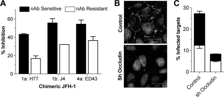
Hepatitis C virus (HCV) can initiate infection by cell-free particle and cell-cell contact-dependent transmission. In this study we use a novel infectious coculture system to examine these alternative modes of infection. Cell-to-cell transmission is relatively resistant to anti-HCV glycoprotein monoclonal antibodies and polyclonal immunoglobulin isolated from infected individuals, providing an effective strategy for escaping host humoral immune responses. Chimeric viruses expressing the structural proteins representing the seven major HCV genotypes demonstrate neutralizing antibody-resistant cell-to-cell transmission. HCV entry is a multistep process involving numerous receptors. In this study we demonstrate that, in contrast to earlier reports, CD81 and the tight-junction components claudin-1 and occludin are all essential for both cell-free and cell-to-cell viral transmission. However, scavenger receptor BI (SR-BI) has a more prominent role in cell-to-cell transmission of the virus, with SR-BI-specific antibodies and small-molecule inhibitors showing preferential inhibition of this infection route. These observations highlight the importance of targeting host cell receptors, in particular SR-BI, to control viral infection and spread in the liver.


Figures

FIG. 1.
FIG. 1.
Effects of anti-glycoprotein antibodies on HCV H77/JFH cell-free infectivity and coculture transmission. Anti-E2 MAbs 9/27, 3/11, and 11/20 (A) and pooled IgG isolated from 6 HCV-infected individuals (B) were titrated in an HCV strain H77/JFH coculture assay. Coculture transmission (open bars), expressed as the percentage of target cells that were infected (left y axis), and the infectivity of cell-free virus (filled bars), expressed as focus-forming units per milliliter (right y axis), were measured. All titrations were performed in duplicate. Error bars indicate standard deviations. The data shown are representative of 3 experiments.
FIG. 2.
FIG. 2.
nAb-resistant HCV transmission requires cell contact and infectious HCV particles. (A) The standard H77/JFH coculture assay was modified either by performing the coculture at a low (0.25×) seeding density or by seeding the target and producer cells on opposing faces of a culture well and transwell insert. In both cases, a 1:1 target/producer ratio was maintained. The frequency of infected target cells in the absence (nAb sensitive) or presence (nAb resistant) of MAb 9/27 at 4 μg/ml is shown. The experiments were performed in duplicate, and error bars indicate standard deviations. The data set is representative of 4 experiments. (B) J6/JFH and J6/JFH del B RNA were electroporated into Huh-7.5 cells. After 72 h, the cells were labeled with CMFDA, and NS5A expression was measured either immediately (T = 0 h) (left) or 48 h after coculture with Huh-7.5 target cells (T = 48 h) (right).
FIG. 3.
FIG. 3.
Diverse HCVcc transmission. Huh-7.5 cells were electroporated with a panel of chimeric JFH-1 HCV RNAs. At 72 h postelectroporation, the cells were labeled with CMFDA and were cocultured with Huh-7.5 target cells in the presence of control or cross-reactive pooled patient HCV+ IgG at 300 μg/ml for 48 h. Extracellular medium was collected. (A) The levels of infectious virus were quantified. (B) The percentage of neutralization by patient HCV+ IgG was determined. (C) The stacked histograms display nAb-resistant cell-to-cell transmission (open portions of bars) and nAb-sensitive transmission (filled portions of bars) for each virus. The infecting genotypes and strain designations are shown at the bottom. All treatments were performed in duplicate, and error bars indicate standard deviations. The data set is representative of 3 experiments.
FIG. 4.
FIG. 4.
HCV coculture transmission is CD81 dependent. (A) Anti-CD81 inhibition of H77/JFH coculture transmission. The stacked histogram displays nAb-resistant cell-to-cell (open portion of bar) and nAb-sensitive (filled portion of bar) transmission in the presence of a control MAb or one of five anti-CD81 MAbs (5 μg/ml) specific for distinct epitopes. (B) Anti-CD81 MAb 2.s131 was titrated in a standard H77/JFH coculture assay, and nAb-sensitive (▪) and -resistant (▵) transmission is shown. All treatments were performed in duplicate, and error bars indicate standard deviations. (C) H77/JFH- and SA13/JFH-infected producer cells were cocultured with Huh-7 Lunet cells or with cells transduced to express human CD81 with or without anti-CD81 (10 μg/ml MAb 2.s131). Representative flow cytometry plots are shown, and the percentages of target cells infected are given in the upper left quadrants. (D) To corroborate the infectious coculture assay, cells were fixed, stained for NS5A, and imaged by confocal microscopy. HCV-infected target cells (red), uninfected producer cells (green), and infected producer cells (orange) are clearly visible, with cell nuclei shown in gray. White arrowheads indicate rare SA13/JFH-infected Lunet target cells. (E) Flow cytometric analysis of CD81 expression in parental Huh-7 Lunet cells and those transduced to express human CD81. Max, maximum.
FIG. 5.
FIG. 5.
Role for claudin-1 and occludin in HCV coculture transmission. Infectious coculture assays were performed with chimeric HCV-infected producer cells bearing the structural proteins of genotypes 1a (H77), 1b (J4), and 4a (ED43). (A) Target cells were incubated with polyclonal rat anti-claudin-1 serum (dilution, 1/100) prior to coculture. Inhibition of nAb-sensitive (filled bars) and nAb-resistant (open bars) transmission is shown. (B) Huh-7.5 cells were transduced with irrelevant lentiviruses or lentiviruses expressing occludin shRNA (sh occludin). One hundred twenty hours later, the cells were fixed, permeabilized, and stained for occludin expression. (C) Huh-7.5 target cells either transduced with irrelevant control shRNA or silenced with occludin shRNA were cocultured with H77/JFH-infected producer cells, and the frequency of nAb-sensitive (filled portions of bars) and nAb-resistant (open portions of bars) transmission was quantified by flow cytometry. All treatments were performed in duplicate; error bars indicate standard deviations; and the data set is representative of two experiments.
FIG. 6.
FIG. 6.
HCV coculture transmission is SR-BI dependent. (A) An anti-SR-BI MAb was titrated in a standard H77/JFH coculture assay. Inhibition of nAb-sensitive (▪) and nAb-resistant (▵) infection is shown. (B) JFH-1- and JFH-1 G451R-infected producer cells were cocultured with Huh-7.5 target cells in the presence or absence of neutralizing patient IgG (300 μg/ml). The stacked histogram displays nAb-resistant cell-to-cell transmission (open portions of bars) and nAb-sensitive transmission (filled portions of bars) for each virus. (C) Effect of an anti-SR-BI MAb (1 μg/ml) on JFH-1 and JFH-1 G451 nAb-resistant (open bars) and -sensitive (filled bars) transmission. (D) Parental Huh-7.5 cells (open bars) or Huh-7.5 cells overexpressing SR-BI (filled bars) were infected with JFH-1 or JFH-1 G451R for 72 h in a standard cell-free infectious assay. HCV-positive cells were enumerated by immunofluorescent microscopy, allowing quantification of infected-cell focus size. The histograms display the percentages of total infection residing in small (1 to 2 cells), medium (3 to 10 cells), large (11 to 30 cells), or very large (31 to 100 cells) infected-cell foci. (E) H77/JFH- and JFH-1-infected producer cells were cocultured with parental Huh-7.5 cells or with Huh-7.5 cells transduced to overexpress SR-BI (≫), in the presence or absence of neutralizing patient IgG (300 μg/ml), and infected target cells were quantified. H77/JFH and JFH-1 nAb-resistant infection of cells overexpressing SR-BI was significantly increased (P, 0.0096 and 0.0037, respectively). (F) Effects of the SR-BI-specific small molecules ITX5061 and ITX7650 (1 μM) on H77/JFH nAb-sensitive (filled bars) and -resistant (open bars) transmission. Treatments were performed in duplicate, and error bars indicate standard deviations.

Similar articles

Cited by

References

    1. Acton, S. L., K. F. Kozarsky, and A. Rigotti. 1999. The HDL receptor SR-BI: a new therapeutic target for atherosclerosis? Mol. Med. Today 5:518-524. - PubMed
    1. André, P., F. Komurian-Pradel, S. Deforges, M. Perret, J. L. Berland, M. Sodoyer, S. Pol, C. Brechot, G. Paranhos-Baccala, and V. Lotteau. 2002. Characterization of low- and very-low-density hepatitis C virus RNA-containing particles. J. Virol. 76:6919-6928. - PMC - PubMed
    1. Bartosch, B., J. Dubuisson, and F. L. Cosset. 2003. Infectious hepatitis C virus pseudo-particles containing functional E1-E2 envelope protein complexes. J. Exp. Med. 197:633-642. - PMC - PubMed
    1. Bitzegeio, J., D. Bankwitz, K. Hueging, S. Haid, C. Brohm, M. B. Zeisel, E. Herrmann, M. Iken, M. Ott, T. F. Baumert, and T. Pietschmann. 2010. Adaptation of hepatitis C virus to mouse CD81 permits infection of mouse cells in the absence of human entry factors. PLoS Pathog. 6:e1000978. - PMC - PubMed
    1. Burlone, M. E., and A. Budkowska. 2009. Hepatitis C virus cell entry: role of lipoproteins and cellular receptors. J. Gen. Virol. 90:1055-1070. - PubMed

Publication types

MeSH terms