

Blood myeloid dendritic cells from HIV-1-infected individuals display a proapoptotic profile characterized by decreased Bcl-2 levels and by caspase-3+ frequencies that are associated with levels of plasma viremia and T cell activation in an exploratory study
- PMID: 20962079
- PMCID: PMC3014213
- DOI: 10.1128/JVI.01118-10
Blood myeloid dendritic cells from HIV-1-infected individuals display a proapoptotic profile characterized by decreased Bcl-2 levels and by caspase-3+ frequencies that are associated with levels of plasma viremia and T cell activation in an exploratory study
Abstract
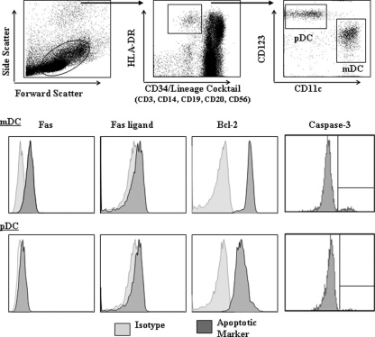
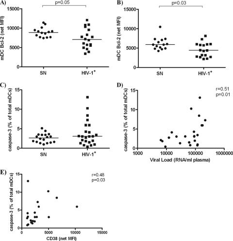
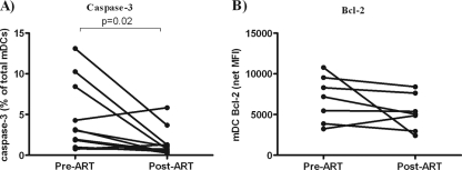
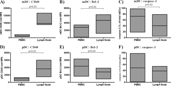
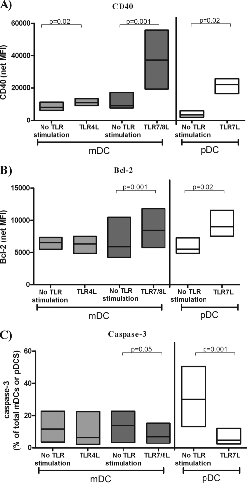
Reduced frequencies of myeloid and plasmacytoid dendritic cell (DC) subsets (mDCs and pDCs, respectively) have been observed in the peripheral blood of HIV-1-infected individuals throughout the course of disease. Accumulation of DCs in lymph nodes (LNs) may partly account for the decreased numbers observed in blood, but increased DC death may also be a contributing factor. We used multiparameter flow cytometry to evaluate pro- and antiapoptotic markers in blood mDCs and pDCs from untreated HIV-1-infected donors, from a subset of infected donors before and after receiving antiretroviral therapy (ART), and from uninfected control donors. Blood mDCs, but not pDCs, from untreated HIV-1-infected donors expressed lower levels of antiapoptotic Bcl-2 than DCs from uninfected donors. A subset of HIV-1-infected donors had elevated frequencies of proapoptotic caspase-3(+) blood mDCs, and positive correlations were observed between caspase-3(+) mDC frequencies and plasma viral load and CD8(+) T-cell activation levels. Caspase-3(+) mDC frequencies, but not mDC Bcl-2 expression, were reduced with viral suppression on ART. Apoptosis markers on DCs in blood and LN samples from a cohort of untreated, HIV-1-infected donors with chronic disease were also evaluated. LN mDCs displayed higher levels of Bcl-2 and lower caspase-3(+) frequencies than did matched blood mDCs. Conversely, LN pDCs expressed lower Bcl-2 levels than their blood counterparts. In summary, blood mDCs from untreated HIV-1-infected subjects displayed a proapoptotic profile that was partially reversed with viral suppression, suggesting that DC death may be a factor contributing to blood DC depletion in the setting of chronic, untreated HIV disease.
Figures
Similar articles
-
Myeloid and lymphoid activation markers in AIDS and non-AIDS presenters.Immunobiology. 2019 Mar;224(2):231-241. doi: 10.1016/j.imbio.2018.11.011. Epub 2018 Nov 28. Immunobiology. 2019. PMID: 30522891
-
Influence of plasma viremia on defects in number and immunophenotype of blood dendritic cell subsets in human immunodeficiency virus 1-infected individuals.J Infect Dis. 2003 Jan 1;187(1):26-37. doi: 10.1086/345957. Epub 2002 Dec 13. J Infect Dis. 2003. PMID: 12508143
-
Antiretroviral therapy partially improves the abnormalities of dendritic cells and lymphoid and myeloid regulatory populations in recently infected HIV patients.Sci Rep. 2019 Aug 12;9(1):11654. doi: 10.1038/s41598-019-48185-2. Sci Rep. 2019. PMID: 31406185 Free PMC article.
-
Dendritic cell dysregulation during HIV-1 infection.Immunol Rev. 2013 Jul;254(1):170-89. doi: 10.1111/imr.12082. Immunol Rev. 2013. PMID: 23772620 Free PMC article. Review.
-
Myeloid dendritic cells in HIV-1 infection.Curr Opin HIV AIDS. 2011 Sep;6(5):379-84. doi: 10.1097/COH.0b013e3283499d63. Curr Opin HIV AIDS. 2011. PMID: 21743323 Free PMC article. Review.
Cited by
-
HIV-1 infection of human intestinal lamina propria CD4+ T cells in vitro is enhanced by exposure to commensal Escherichia coli.J Immunol. 2012 Jul 15;189(2):885-96. doi: 10.4049/jimmunol.1200681. Epub 2012 Jun 11. J Immunol. 2012. PMID: 22689879 Free PMC article.
-
A divergent myeloid dendritic cell response at virus set-point predicts disease outcome in SIV-infected rhesus macaques.J Med Primatol. 2011 Aug;40(4):206-13. doi: 10.1111/j.1600-0684.2011.00484.x. Epub 2011 Jul 1. J Med Primatol. 2011. PMID: 21718317 Free PMC article.
-
Increased Escherichia coli-induced interleukin-23 production by CD16+ monocytes correlates with systemic immune activation in untreated HIV-1-infected individuals.J Virol. 2013 Dec;87(24):13252-62. doi: 10.1128/JVI.01767-13. Epub 2013 Sep 25. J Virol. 2013. PMID: 24067979 Free PMC article.
-
Insights into the apoptotic death of immune cells in sepsis.J Interferon Cytokine Res. 2015 Jan;35(1):17-22. doi: 10.1089/jir.2014.0069. Epub 2014 Jul 9. J Interferon Cytokine Res. 2015. PMID: 25007137 Free PMC article. Review.
-
Differential interleukin-10 (IL-10) and IL-23 production by human blood monocytes and dendritic cells in response to commensal enteric bacteria.Clin Vaccine Immunol. 2012 Aug;19(8):1207-17. doi: 10.1128/CVI.00282-12. Epub 2012 Jun 13. Clin Vaccine Immunol. 2012. PMID: 22695160 Free PMC article.
References
-
- Airo, P., C. Torti, M. C. Uccelli, F. Malacarne, L. Palvarini, G. Carosi, and F. Castelli. 2000. Naive CD4+ T lymphocytes express high levels of Bcl-2 after highly active antiretroviral therapy for HIV infection. AIDS Res. Hum. Retrovir. 16:1805-1807. - PubMed
-
- Ashe, P. C., and M. D. Berry. 2003. Apoptotic signaling cascades. Prog. Neuropsychopharmacol. Biol. Psychiatry. 27:199-214. - PubMed
-
- Balestrieri, E., S. Grelli, C. Matteucci, A. Minutolo, G. d'Ettorre, F. Di Sora, F. Montella, V. Vullo, S. Vella, C. Favalli, B. Macchi, and A. Mastino. 2007. Apoptosis-associated gene expression in HIV-infected patients in response to successful antiretroviral therapy. J. Med. Virol. 79:111-117. - PubMed
-
- Banchereau, J., F. Briere, C. Caux, J. Davoust, S. Lebecque, Y. J. Liu, B. Pulendran, and K. Palucka. 2000. Immunobiology of dendritic cells. Annu. Rev. Immunol. 18:767-811. - PubMed
Publication types
MeSH terms
Substances
Grants and funding
LinkOut - more resources
Full Text Sources
Other Literature Sources
Research Materials
