. 2010 Oct 20;30(42):14059-67.
doi: 10.1523/JNEUROSCI.3883-10.2010.

14-3-3 proteins regulate protein kinase a activity to modulate growth cone turning responses

Affiliations

14-3-3 proteins regulate protein kinase a activity to modulate growth cone turning responses

Christopher B Kent et al. J Neurosci. .

Abstract

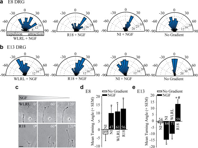
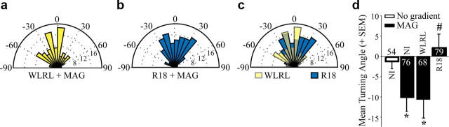
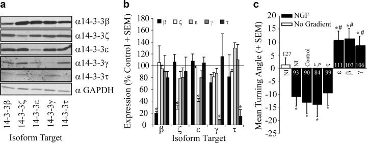
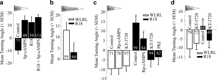
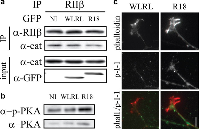
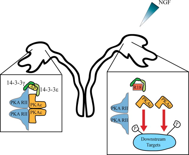
Growth cones regulate the speed and direction of neuronal outgrowth during development and regeneration. How the growth cone spatially and temporally regulates signals from guidance cues is poorly understood. Through a proteomic analysis of purified growth cones we identified isoforms of the 14-3-3 family of adaptor proteins as major constituents of the growth cone. Disruption of 14-3-3 via the R18 antagonist or knockdown of individual 14-3-3 isoforms switches nerve growth factor- and myelin-associated glycoprotein-dependent repulsion to attraction in embryonic day 13 chick and postnatal day 5 rat DRG neurons. These effects are reminiscent of switching responses observed in response to elevated cAMP. Intriguingly, R18-dependent switching is blocked by inhibitors of protein kinase A (PKA), suggesting that 14-3-3 proteins regulate PKA. Consistently, 14-3-3 proteins interact with PKA and R18 activates PKA by dissociating its regulatory and catalytic subunits. Thus, 14-3-3 heterodimers regulate the PKA holoenzyme and this activity plays a critical role in modulating neuronal responses to repellent cues.


Figures

Figure 1.
Figure 1.
14-3-3 proteins are present in growth cones. Immunofluorescence staining of growth cones with isoform-specific 14-3-3 antibodies. For each antibody the immunofluorescence signal is lost by preadsorbing the antibody with isoform-specific blocking peptide (BP). RGC, retinal ganglion cell. Scale bar, 5 μm.
Figure 2.
Figure 2.
The 14-3-3 antagonist R18 converts NGF-dependent repulsion to attraction in E13 chick DRG neurons. a, b, Rose histograms illustrating turning responses of E8 (a) or E13 (b) chick DRG neurons in response to an NGF gradient in a Dunn chamber turning assay (50 ng/ml NGF in outer chamber). Responses of individual neurons are clustered in 10° bins and the percentage of total neurons per bin is represented by the radius of each segment. Nontransduced neurons (NI) or neurons transduced with HSV-WLRL-GFP or HSV-R18-GFP for 12–16 h before the turning assay were analyzed. c, Phase images of representative E13 chick DRG growth cones transduced with WLRL or R18 and exposed to an NGF gradient in the Dunn chamber turning assay. Scale bar, 20 μm. d, e, Mean turning angles of E8 (d) or E13 (e) chick DRG neurons in the absence of a cue (No Gradient) or in response to 50 ng/ml NGF. Numbers within the bars indicate the number of growth cones measured over at least three independent experiments. Statistics were performed by one-way ANOVA with Tukey's post hoc test. *p < 0.05 compared with no gradient control; #p < 0.05 compared with the NGF-dependent repellent response in WLRL-transduced neurons.
Figure 3.
Figure 3.
The 14-3-3 antagonist R18 blocks MAG-dependent repulsion of E13 chick DRG neurons. a, b, Rose histograms illustrate turning responses of E13 chick DRG neurons in response to a MAG-Fc gradient (200 ng/ml in outer chamber) in a Dunn chamber turning assay. Neurons were transduced with HSV-WLRL (a) or HSV-R18 (b) for 12–16 h before the turning assay. c, An overlay of the rose histograms presented in a and b illustrating a large population of R18-positive neurons with an attractive turning response to a MAG gradient. d, Mean turning angles of noninfected, WLRL- or R18-transduced E13 chick DRG neurons in the absence of a gradient or in response to MAG. Numbers within the bars indicate the number of growth cones measured over at least three independent experiments. Statistics were performed by one-way ANOVA with Tukey's post hoc test. *p < 0.05 compared with no gradient control; #p < 0.05 compared with the MAG-dependent repellent response in WLRL-transduced neurons.
Figure 4.
Figure 4.
Loss of 14-3-3ε, β or γ converts NGF-dependent repulsion to attraction in P5 rat DRG neurons. a, Lysates from P5 rat DRG neurons transduced with lentiviruses for knockdown of individual 14-3-3 isoforms and analyzed by Western blot with anti-GAPDH or anti-14-3-3 isoform-specific antibodies. The GAPDH blot shown is from the 14-3-3β gel and is representative of the equal loading achieved in these lysates. b, Expression of individual isoforms quantified by densitometry from Western blots of P5 rat DRG lysates transduced with lentiviruses for knockdown of individual isoforms. Densities were normalized to GAPDH levels for each blot. The mean expression of each isoform is expressed as a percentage of control from 3 independent experiments (+SEM). *p < 0.05 compared with expression in control miRNA-transduced neurons. c, Mean turning angles of P5 rat DRG neurons in the absence of a cue (No Gradient) or in response to 100 ng/ml NGF. Neurons were nontransduced (NI) or were transduced with lentiviruses for expression of a nontargeting control sequence or sequences for knockdown of individual 14-3-3 isoforms. Numbers within the bars indicate the number of growth cones measured over at least three independent experiments. Statistics were performed by one-way ANOVA with Tukey's post hoc test. *p < 0.05 compared with no gradient control; #p < 0.05 compared with the NGF-dependent repellent response in control shRNAmir-transduced neurons.
Figure 5.
Figure 5.
14-3-3 proteins regulate growth cone turning responses through PKA. a–d, Mean turning angles of E13 chick DRG neurons in response to gradients of NGF (50 ng/ml), Sp-cAMPS (20 μm), or MAG-Fc (200 ng/ml). Neurons were transduced with HSV-WLRL-GFP or HSV-R18-GFP. Where indicated, neurons were treated with a bath application of 20 μm Sp-cAMPS, 20 μm Rp-cAMPS, 200 nm KT-5720, or 20 μm myristoylated PKI for 60 min before the turning analysis. Numbers within the bars indicate the number of growth cones measured over at least three independent experiments. Statistics for a, c, and d were performed by two-way ANOVA with Bonferroni post hoc tests. Statistics for b and PKI treatment were performed with unpaired Student's t test. *p < 0.05 compared with the WLRL-transduced control for each condition.
Figure 6.
Figure 6.
14-3-3 proteins bind PKA. a, b, 293T cells were cotransfected with V5-tagged RIIα or RIIβ and myc-tagged 14-3-3 constructs and GFP-WLRL or GFP-R18 and then subjected to immunoprecipitation with anti-V5 (a) or anti-myc (b) antibody. Cell lysates and immunoprecipitates were separated by SDS-PAGE and analyzed by Western blotting with anti-V5 and anti-myc antibodies. c, Cell lysates from P5 rat DRG neurons were subjected to immunoprecipitation with control IgG or anti-RIIβ antibody and cell lysates and immunoprecipitates were analyzed by Western blot with anti-14-3-3γ and anti-RIIβ antibodies.
Figure 7.
Figure 7.
14-3-3 proteins regulate the stability and activity of PKA holoenzyme. a, PC12 cells were transduced with HSV-WLRL or HSV-R18. Following transduction, cell lysates were subjected to immunoprecipitation with anti-RIIβ antibody. Cell lysates and immunoprecipitates were analyzed by Western blot with anti-RIIβ, anti-PKA catalytic subunit (cat) and anti-GFP antibodies. b, P5 rat DRGs were transduced with HSV-WLRL or HSV-R18. Cell lysates were analyzed by Western blot for levels of phosphorylated PKA catalytic subunit and total levels of PKA catalytic subunit. c, Immunofluorescence of P5 rat DRG growth cones transduced with HSV-WLRL or HSV-R18 and stained with an anti-phospho-I-1 antibody and rhodamine-phalloidin. Scale bar, 10 μm.
Figure 8.
Figure 8.
Model for 14-3-3 regulation of PKA in growth cone turning response. In P5 rat DRG growth cones, 14-3-3γ/ε heterodimers (shown in green) bind to PKA directly or through an adaptor protein (AP) and downregulate the activity of the PKA holoenzyme, resulting in a repellant response to a gradient of NGF (left). In the presence of the R18 peptide (red square), 14-3-3 binding is disrupted, leading to a dissociation of the active catalytic subunits of PKA (orange) from the regulatory subunits (blue) and an increase in PKA phosphorylation of downstream targets. This switches the response of the growth cone from repulsion to attraction (right).

Similar articles

Cited by

References

    1. Berruti G. A novel rap1/B-Raf/14-3-3 theta protein complex is formed in vivo during the morphogenetic differentiation of postmeiotic male germ cells. Exp Cell Res. 2000;257:172–179. - PubMed
    1. Bouchard JF, Moore SW, Tritsch NX, Roux PP, Shekarabi M, Barker PA, Kennedy TE. Protein kinase A activation promotes plasma membrane insertion of DCC from an intracellular pool: a novel mechanism regulating commissural axon extension. J Neurosci. 2004;24:3040–3050. - PMC - PubMed
    1. Bridges D, Moorhead GB. 14-3-3 proteins: a number of functions for a numbered protein. Sci STKE. 2005;2005:re10. - PubMed
    1. Cai D, Qiu J, Cao Z, McAtee M, Bregman BS, Filbin MT. Neuronal cyclic AMP controls the developmental loss in ability of axons to regenerate. J Neurosci. 2001;21:4731–4739. - PMC - PubMed
    1. Clandinin TR, Zipursky SL. Making connections in the fly visual system. Neuron. 2002;35:827–841. - PubMed

Publication types

MeSH terms

Substances

LinkOut - more resources