. 2010 Dec;21(24):4387-99.
doi: 10.1091/mbc.E10-06-0489. Epub 2010 Oct 20.

Podoplanin associates with CD44 to promote directional cell migration

Affiliations

Podoplanin associates with CD44 to promote directional cell migration

Ester Martín-Villar et al. Mol Biol Cell. 2010 Dec.

Abstract

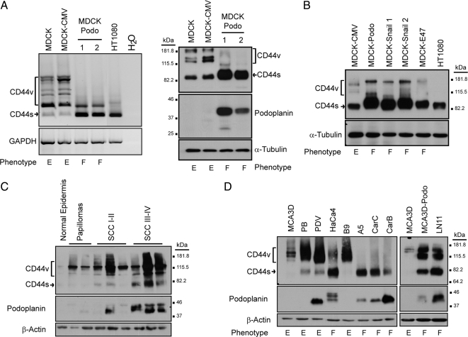
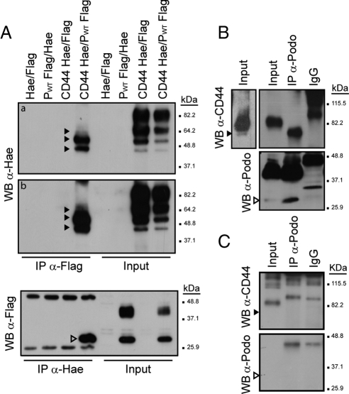
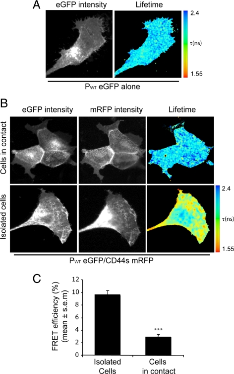
Podoplanin is a transmembrane glycoprotein up-regulated in different human tumors, especially those derived from squamous stratified epithelia (SCCs). Its expression in tumor cells is linked to increased cell migration and invasiveness; however, the mechanisms underlying this process remain poorly understood. Here we report that CD44, the major hyaluronan (HA) receptor, is a novel partner for podoplanin. Expression of the CD44 standard isoform (CD44s) is coordinately up-regulated together with that of podoplanin during progression to highly aggressive SCCs in a mouse skin model of carcinogenesis, and during epithelial-mesenchymal transition (EMT). In carcinoma cells, CD44 and podoplanin colocalize at cell surface protrusions. Moreover, CD44 recruitment promoted by HA-coated beads or cross-linking with a specific CD44 antibody induced corecruitment of podoplanin. Podoplanin-CD44s interaction was demonstrated both by coimmunoprecipitation experiments and, in vivo, by fluorescence resonance energy transfer/fluorescence lifetime imaging microscopy (FRET/FLIM), the later confirming its association on the plasma membrane of cells with a migratory phenotype. Importantly, we also show that podoplanin promotes directional persistence of motility in epithelial cells, a feature that requires CD44, and that both molecules cooperate to promote directional migration in SCC cells. Our results support a role for CD44-podoplanin interaction in driving tumor cell migration during malignancy.


Figures

Figure 1.
Figure 1.
Podoplanin and CD44 expression during EMT and mouse skin carcinogenesis. (A) Podoplanin-promoted EMT in MDCK cells is associated with induction of CD44s expression. The phenotype of the cell lines is indicated: E, epithelial; F, fibroblastic. The morphology and characterization of these cells is shown in Supplemental Figure S1 and in Martin-Villar et al. (2006). The fibrosarcoma HT1080 cell line was used as a positive control for CD44s expression. Left and right panels show, respectively, the expression of CD44 transcripts (RT-PCR) and proteins (Western blot). (B) CD44 protein expression in MDCK cells that underwent EMT by transfection of E-cadherin repressors Snail 1, 2, and E47. (C) CD44 and podoplanin protein expression in mouse normal epidermis and skin tumors induced by two-stage carcinogenesis. All squamous cell carcinomas (SCCs) were excised at 43 wk post-initiation. SCCI-II, well to moderately differentiated; SCCIII-IV, poorly differentiated. (D) CD44 and podoplanin protein expression in mouse epidermal cell lines (see table S1 for further information of the cells lines). MCA3D keratinocytes forced to express podoplanin (MCA3D-Podo) undergo EMT. CD44s, CD44 standard isoform; CD44v, variant CD44 isoforms. GAPDH and α-tubulin/β-actin were used as loading controls for RNA and protein, respectively.
Figure 2.
Figure 2.
Podoplanin and CD44 colocalize at cell-surface protrusions. (A) Confocal microscopy showing localization of endogenous podoplanin and CD44 in HN5 oral carcinoma cells, and in MDCK cells stably transfected with podoplanin-eGFP (PWT eGFP). (B) Recruitment of both podoplanin and CD44 by HA-coated beads (arrowheads). MDCK-PWT eGFP cells plated onto coverslips were incubated with 5-μm beads coated with BSA or HA for 15 min. Cells were then processed for immunofluorescence to detect CD44. (C) Ab-induced clustering of CD44 (HP2/9 mAb, red) induced corecruitment of PWT GFP (green) to CD44 patches (arrowheads) in HN5 cells. CD44 staining (blue) was performed using a different CD44 mAb (IM7) to visualize total distribution of CD44. Mouse IgGs were used as a negative control. Bars, 10 μm.
Figure 3.
Figure 3.
Podoplanin coimmunoprecipitates with CD44s. (A) HEK293T were transiently cotransfected with Flag-tagged human podoplanin or Hae-tagged human CD44s, as indicated. After 24 h of transfection, total lysates were immunoprecipitated with anti-Hae or anti-Flag Abs. Immunoprecipitates were electrophoresed by 10% SDS-PAGE and immunoblotted with anti-Flag or anti-Hae Abs, respectively. Panels a and b represent different exposure times of the same immunoblot. (B) Coimmunoprecipitation of endogenous CD44 and podoplanin in the mouse carcinoma cell line CarC. Lysates were immunoprecipitated with either a mAb specific for mouse podoplanin (PA2.26) or rat IgG as a control. The presence of podoplanin and CD44s in the precipitate was determined with anti-podoplanin PA2.26 and anti-CD44 IM7 mAbs, respectively. A longer exposure time of the input showing the occurrence of a ∼70 kDa CD44s form in CarC lysates is shown in the left panel. (C) Coimmunoprecipitation assay in HaCaT keratinocytes. Note that HaCaT cells do not express podoplanin and were used as negative control for the coimmunoprecipitation assays depicted in panel B. Arrowheads indicate incompletely glycosylated forms of CD44s (black arrowheads) and podoplanin (open arrowheads).
Figure 4.
Figure 4.
Podoplanin–CD44s complexes at the plasma membrane are up-regulated in cells with a migratory phenotype. (A–C) Multiphoton FLIM was used to image FRET between PWT eGFP (donor) and CD44s-mRFP (acceptor) in MDCK cells. The images show the eGFP multiphoton intensity image and (where appropriate) the corresponding wide-field CCD camera image of mRFP expression. Lifetime images mapping spatial FRET across the cells are depicted using a pseudocolor scale (blue, normal eGFP lifetime; red, FRET). (A) Control MDCK cells expressing PWT eGFP alone demonstrated a normal GFP lifetime (τ in ns) in the absence of acceptor. (B) Cells coexpressing PWT eGFP and CD44s-mRFP display a localized shortening of the eGFP fluorescence lifetime, which is demonstrated by red in the pseudocolor scale. Note that although colocalization between PWT eGFP and CD44s–mRFP was always detected, FRET was recorded mainly in isolated cells (i.e., those that had detached from their neighbors). (C) Bar graph representing average FRET efficiency of 14 cells for each condition over three independent experiments. The Student's t test was used to evaluate statistical significance between different populations of data. ***p < 0.0005.
Figure 5.
Figure 5.
Podoplanin–CD44s interaction is not mediated by ERM proteins. (A) Schematic representation of podoplanin and CD44s fusion constructs used for coimmunoprecipitation and FRET/FLIM assays. SP, signal peptide; Ec, ectodomain; TM, transmembrane domain; CT, cytoplasmic tail; QN N, positive charged residues (RK.R) in podoplanin juxtamembrane domain were substituted by uncharged polar amino acids (QN.N) in order to impair podoplanin binding to ERM proteins (Martin-Villar et al., 2006). (B) MDCK cells were cotransfected with CD44s mRFP and podoplanin eGFP mutant constructs, and cells were then imaged by FLIM to detect FRET as depicted in Figure 4. Images show the eGFP multiphoton intensity image and (where appropriate) the corresponding wide-field CCD camera image of the mRFP expression. Control MDCK cells expressing PWT, PΔCT, or PQNN eGFP alone showed a normal GFP lifetime (τ in ns) in the absence of acceptor (CD44s mRFP), while cells coexpressing CD44s mRFP and podoplanin mutant constructs displayed a localized shortening of the eGFP fluorescence lifetime. (C) The bar graph represents average FRET efficiency of 15 cells over three independent experiments. (D) Coimmunoprecipitation assays performed in HEK293T cells coexpressing CD44s Hae and podoplanin eGFP mutant constructs. Total cell lysates were immunoprecipitated with an anti-GFP Ab-conjugated resin as described in Materials and Methods. The coimmunoprecipitated products were detected with an anti-Hae Ab.
Figure 6.
Figure 6.
Podoplanin-induced migration and directionality in MDCK cells requires CD44. (A) Expression levels of podoplanin (PWT eGFP) and CD44 after PWT eGFP and CD44 siRNA coexpression. β-actin was used as a loading control. Arrowheads indicate fully (black arrowheads) and incompletely glycosylated (open arrowheads) forms of PWT eGFP. (B) Transwell migration assay. Bar graph representing percentage of migrating cells per total number of cells. Results are representative of three independent experiments performed in duplicates. (C) Representative migration tracks of MDCK cells expressing PWT eGFP in the presence or absence of CD44 (n = 30). (D) Average persistence and speed of migration derived from the tracks depicted in C. n = 40–85 cells per bar. Asterisks indicate significant differences in a Student's t test. **p < 0.005, *p < 0.05.
Figure 7.
Figure 7.
Knockdown of CD44 and podoplanin in oral carcinoma HN5 cells affects cell spreading. (A) Expression levels of CD44 and podoplanin in single and double knockdown cells by Western blot analysis. The expression of GAPDH was used as a loading control. (B) Immunofluorescence detection of F-actin and microtubules in single and double knockdown cells. Cells were double stained for F-actin (red) with phalloidin and microtubules (green) with a specific anti-tubulin mAb. Note the extremely disorganized leading edge of podoplanin-deficient cells and double knockdown cells (arrowheads) compared with control cells. (C) Graphical representation of the cell spread areas quantified using Image J software as described in Materials and Methods (n = 40–50 cells per bar). P values were obtained using a one-way analysis of variance (ANOVA). *p < 0.05; **p < 0.005; ***p < 0.0005. Bars, 10 μm.
Figure 8.
Figure 8.
Knockdown of CD44 and podoplanin in oral carcinoma HN5 cells impairs directional migration during wound healing. (A) Analysis of the migration pattern at the wound edge of podoplanin and CD44 knocked-down HN5 cells. The migration paths of representative cells taken from the wound edge are indicated on an overlay image from the initiation of the imaging (0 h). Images of the wounds at 6 h after the incision was made are shown in the middle panel. Trace of the movement of multiple cells along the wound edge (n = 30) are shown in the graphs of the right panel. (B) Quantification of the wound closure after 18 h. (C) Average persistence of migration (directionality) from the tracks depicted in A. An animate and complete (18h) sequence of these data are shown in Supplemental Movies 1 and 2. P values were obtained using one-way analysis of variance (ANOVA). *p < 0.05; **p < 0.005, ***p < 0.0005.

References

    1. Akhurst R. J., Balmain A. Genetic events and the role of TGF beta in epithelial tumour progression. J. Pathol. 1999;187:82–90. - PubMed
    1. Allen W. E., Zicha D., Ridley A. J., Jones G. E. A role for Cdc42 in macrophage chemotaxis. J. Cell Biol. 1998;141:1147–1157. - PMC - PubMed
    1. Atsumi N., Ishii G., Kojima M., Sanada M., Fujii S., Ochiai A. Podoplanin, a novel marker of tumor-initiating cells in human squamous cell carcinoma A431. Biochem. Biophys. Res. Commun. 2008;373:36–41. - PubMed
    1. Burns P. A., Kemp C. J., Gannon J. V., Lane D. P., Bremner R., Balmain A. Loss of heterozygosity and mutational alterations of the p53 gene in skin tumours of interspecific hybrid mice. Oncogene. 1991;6:2363–2369. - PubMed
    1. Camp R. L., Kraus T. A., Pure E. Variations in the cytoskeletal interaction and posttranslational modification of the CD44 homing receptor in macrophages. J. Cell Biol. 1991;115:1283–1292. - PMC - PubMed

Publication types

MeSH terms