

Visualization of lymphatic vessels by Prox1-promoter directed GFP reporter in a bacterial artificial chromosome-based transgenic mouse
- PMID: 20962325
- PMCID: PMC3037757
- DOI: 10.1182/blood-2010-07-298562
Visualization of lymphatic vessels by Prox1-promoter directed GFP reporter in a bacterial artificial chromosome-based transgenic mouse
Abstract
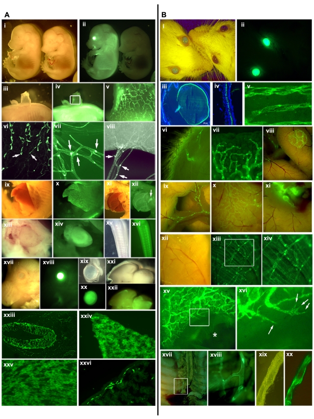
Although the blood vessel-specific fluorescent transgenic mouse has been an excellent tool to study vasculogenesis and angiogenesis, a lymphatic-specific fluorescent mouse model has not been established to date. Here we report a transgenic animal model that expresses the green fluorescent protein under the promoter of Prox1, a master control gene in lymphatic development. Generated using an approximately 200-kb-long bacterial artificial chromosome harboring the entire Prox1 gene, this Prox1-green fluorescent protein mouse was found to faithfully recapitulate the expression pattern of the Prox1 gene in lymphatic endothelial cells and other Prox1-expressing organs, and enabled us to conveniently visualize detailed structure and morphology of lymphatic vessels and networks throughout development. Our data demonstrate that this novel transgenic mouse can be extremely useful for detection, imaging, and isolation of lymphatic vessels and monitoring wound-associated lymphangiogenesis. Together, this Prox1-green fluorescent protein transgenic mouse will be a great tool for the lymphatic research.
Figures

Similar articles
-
Development and Characterization of A Novel Prox1-EGFP Lymphatic and Schlemm's Canal Reporter Rat.Sci Rep. 2017 Jul 17;7(1):5577. doi: 10.1038/s41598-017-06031-3. Sci Rep. 2017. PMID: 28717161 Free PMC article.
-
ProxTom lymphatic vessel reporter mice reveal Prox1 expression in the adrenal medulla, megakaryocytes, and platelets.Am J Pathol. 2012 Apr;180(4):1715-25. doi: 10.1016/j.ajpath.2011.12.026. Epub 2012 Feb 4. Am J Pathol. 2012. PMID: 22310467 Free PMC article.
-
Lymphatic vessel function in head and neck inflammation.Lymphat Res Biol. 2013 Sep;11(3):187-92. doi: 10.1089/lrb.2013.0013. Lymphat Res Biol. 2013. PMID: 24044758 Free PMC article.
-
Intravital two-photon microscopy of lymphatic vessel development and function using a transgenic Prox1 promoter-directed mOrange2 reporter mouse.Biochem Soc Trans. 2011 Dec;39(6):1674-81. doi: 10.1042/BST20110722. Biochem Soc Trans. 2011. PMID: 22103506 Review.
-
Fluorescent reporter transgenic mice for in vivo live imaging of angiogenesis and lymphangiogenesis.Angiogenesis. 2018 Nov;21(4):677-698. doi: 10.1007/s10456-018-9629-2. Epub 2018 Jul 3. Angiogenesis. 2018. PMID: 29971641 Free PMC article. Review.
Cited by
-
Penile cavernous sinusoids are Prox1-positive hybrid vessels.Vasc Biol. 2024 Jan 11;6(1):e230014. doi: 10.1530/VB-23-0014. Print 2024 Jan 1. Vasc Biol. 2024. PMID: 38051669 Free PMC article.
-
Exosomes as a Communication Tool Between the Lymphatic System and Bladder Cancer.Int Neurourol J. 2018 Sep;22(3):220-224. doi: 10.5213/inj.1836186.093. Epub 2018 Sep 28. Int Neurourol J. 2018. PMID: 30286586 Free PMC article. No abstract available.
-
c-JUN-mediated transcriptional responses in lymphatic endothelial cells are required for lung fluid clearance at birth.Proc Natl Acad Sci U S A. 2023 Jan 10;120(2):e2215449120. doi: 10.1073/pnas.2215449120. Epub 2023 Jan 3. Proc Natl Acad Sci U S A. 2023. PMID: 36595691 Free PMC article.
-
Leukocyte Trafficking in Lymphatic Vessels.Cold Spring Harb Perspect Med. 2022 Oct 3;12(10):a041186. doi: 10.1101/cshperspect.a041186. Cold Spring Harb Perspect Med. 2022. PMID: 35379657 Free PMC article. Review.
-
Initial afferent lymphatic vessels controlling outbound leukocyte traffic from skin to lymph nodes.Front Immunol. 2013 Dec 9;4:433. doi: 10.3389/fimmu.2013.00433. Front Immunol. 2013. PMID: 24368908 Free PMC article. Review.
References
-
- Motoike T, Loughna S, Perens E, et al. Universal GFP reporter for the study of vascular development. Genesis. 2000;28(2):75–81. - PubMed
-
- Wigle JT, Oliver G. Prox1 function is required for the development of the murine lymphatic system. Cell. 1999;98(6):769–778. - PubMed
-
- Gong S, Zheng C, Doughty ML, et al. A gene expression atlas of the central nervous system based on bacterial artificial chromosomes. Nature. 2003;425(6961):917–925. - PubMed
-
- Petrova TV, Nykanen A, Norrmen C, et al. Transcription factor PROX1 induces colon cancer progression by promoting the transition from benign to highly dysplastic phenotype. Cancer Cell. 2008;13(5):407–419. - PubMed
Publication types
MeSH terms
Substances
Grants and funding
LinkOut - more resources
Full Text Sources
Other Literature Sources
Molecular Biology Databases
