

Deciphering the human platelet sheddome
- PMID: 20962327
- PMCID: PMC3037762
- DOI: 10.1182/blood-2010-05-283838
Deciphering the human platelet sheddome
Abstract
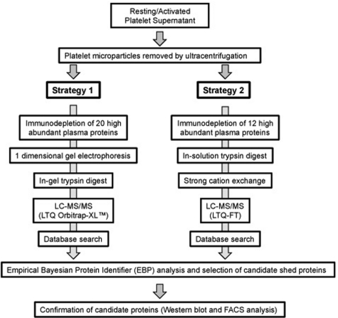
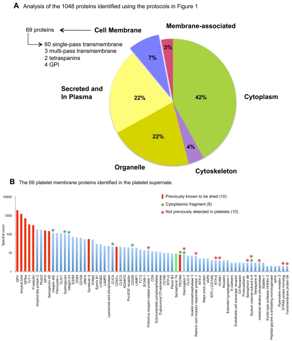
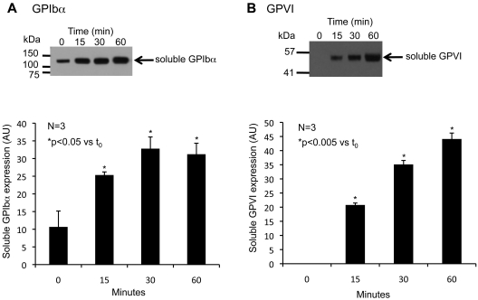
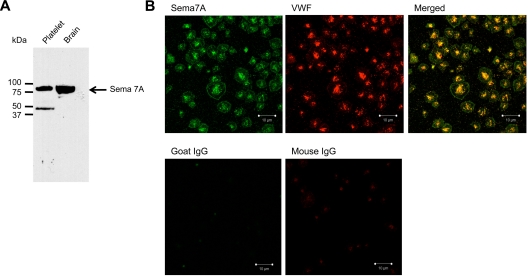
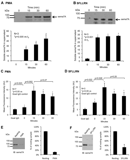
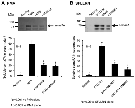
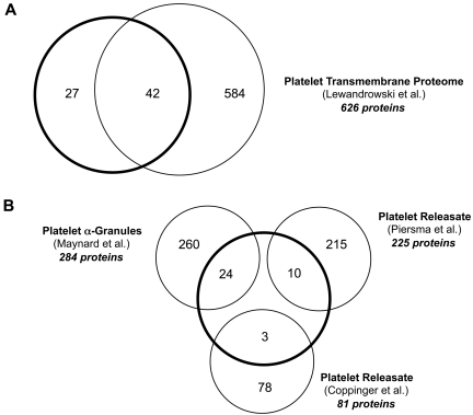
Activated platelets shed surface proteins, potentially modifying platelet function as well as providing a source of bioactive fragments. Previous studies have identified several constituents of the platelet sheddome, but the full extent of shedding is unknown. Here we have taken a global approach, analyzing protein fragments in the supernate of activated platelets using mass spectroscopy and looking for proteins originating from platelet membranes. After removing plasma proteins and microparticles, 1048 proteins were identified, including 69 membrane proteins. Nearly all of the membrane proteins had been detected previously, but only 10 had been shown to be shed in platelets. The remaining 59 are candidates subject to confirmation. Based on spectral counts, protein representation in the sheddome varies considerably. As proof of principle, we validated one of the less frequently detected proteins, semaphorin 7A, which had not previously been identified in platelets. Surface expression, cleavage, and shedding of semaphorin 7A were demonstrated, as was its association with α-granules. Finally, cleavage of semaphorin 7A and 12 other proteins was substantially reduced by an inhibitor of ADAM17, a known sheddase. These results define a subset of membrane proteins as sheddome candidates, forming the basis for further studies examining the impact of ectodomain shedding on platelet function.
Figures
References
-
- Andrews RK, Lopez JA, Berndt MC. Molecular mechanisms of platelet adhesion and activation. Int J Biochem Cell Biol. 1997;29(1):91–105. - PubMed
-
- Varga-Szabo D, Pleines I, Nieswandt B. Cell adhesion mechanisms in platelets. Arterioscler Thromb Vasc Biol. 2008;28(3):403–412. - PubMed
-
- Wei AH, Schoenwaelder SM, Andrews RK, Jackson SP. New insights into the haemostatic function of platelets. Br J Haematol. 2009;147(4):415–430. - PubMed
-
- Baurand A, Eckly A, Hechler B, et al. Differential regulation and relocalization of the platelet P2Y receptors after activation: a way to avoid loss of hemostatic properties? Mol Pharmacol. 2005;67(3):721–733. - PubMed
-
- Schober JM, Lam SC, Wencel-Drake JD. Effect of cellular and receptor activation on the extent of integrin alphaIIbbeta3 internalization. J Thromb Haemost. 2003;1(11):2404–2410. - PubMed
Publication types
MeSH terms
Substances
Grants and funding
LinkOut - more resources
Full Text Sources
Research Materials
Miscellaneous
