. 2010 Nov 4;468(7320):93-7.
doi: 10.1038/nature09494. Epub 2010 Oct 20.

Antibodies to human serum amyloid P component eliminate visceral amyloid deposits

Affiliations

Antibodies to human serum amyloid P component eliminate visceral amyloid deposits

Karl Bodin et al. Nature. .

Abstract

Accumulation of amyloid fibrils in the viscera and connective tissues causes systemic amyloidosis, which is responsible for about one in a thousand deaths in developed countries. Localized amyloid can also have serious consequences; for example, cerebral amyloid angiopathy is an important cause of haemorrhagic stroke. The clinical presentations of amyloidosis are extremely diverse and the diagnosis is rarely made before significant organ damage is present. There is therefore a major unmet need for therapy that safely promotes the clearance of established amyloid deposits. Over 20 different amyloid fibril proteins are responsible for different forms of clinically significant amyloidosis and treatments that substantially reduce the abundance of the respective amyloid fibril precursor proteins can arrest amyloid accumulation. Unfortunately, control of fibril-protein production is not possible in some forms of amyloidosis and in others it is often slow and hazardous. There is no therapy that directly targets amyloid deposits for enhanced clearance. However, all amyloid deposits contain the normal, non-fibrillar plasma glycoprotein, serum amyloid P component (SAP). Here we show that administration of anti-human-SAP antibodies to mice with amyloid deposits containing human SAP triggers a potent, complement-dependent, macrophage-derived giant cell reaction that swiftly removes massive visceral amyloid deposits without adverse effects. Anti-SAP-antibody treatment is clinically feasible because circulating human SAP can be depleted in patients by the bis-d-proline compound CPHPC, thereby enabling injected anti-SAP antibodies to reach residual SAP in the amyloid deposits. The unprecedented capacity of this novel combined therapy to eliminate amyloid deposits should be applicable to all forms of systemic and local amyloidosis.


Figures

Figure 1
Figure 1. Elimination of visceral amyloid in AA amyloidotic mice after treatment with anti-SAP antibody
Systemic AA amyloidosis was established in C57BL/6 SAP deficient mice transgenically expressing human SAP, which were allocated to 3 groups closely matched for age, sex and whole body amyloid load, the latter shown as mean (SD) per cent whole body retention of 125I-human SAP tracer (one way ANOVA comparing the 3 group means, P=0.054). The mice were treated with CPHPC and a single dose of sheep anti-human SAP antibody (group 1, n=31), with CPHPC and the same dose of an irrelevant sheep anti-human antibody (group 2, n=30), or left untreated (group 3, n=32). Amyloid load was determined histologically 28 days later. a, Each point is an individual spleen amyloid score. 0, no amyloid detected; 1, one or more trace specks; 2, marginal zone traces; 3, general marginal zone deposits; 4, heavy marginal zone deposits; 5, heavy marginal zone and extensive interfollicular deposits. Kruskal-Wallis test comparing the 3 groups, P<0.0001. Dunn’s multiple comparison test: 1 vs 2 P<0.001; 1 vs 3 P<0.001; 2 vs 3 not significant P>0.05. b, Congo red stained spleen section showing the pathognomonic amyloid green dichroism, score = 5. c, As in b but with amyloid score = 1; single amyloid speck is arrowed. d, Individual liver amyloid scores. 0, no amyloid detected; 1, trace specks; 2, traces in/around most portal tracts; 3, significant deposits in/around all portal tracts; 4, extensive portal and parenchymal deposits. Kruskal-Wallis test, P<0.0001. Dunn’s multiple comparison test: 1 vs 2 P<0.001; 1 vs 3 P<0.001; 2 vs 3 not significant P>0.05. There were no significant differences in the average spleen or liver amyloid scores between males and females within any of the groups (not shown). e, Congo red stain of liver amyloid, score = 4. f, Liver amyloid score = 1; arrows indicate amyloid specks.
Figure 2
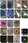
Figure 2. Cellular infiltration and amyloid destruction after administration of anti-SAP antibody
a, Congo red stained spleen section one day after anti-SAP antibody treatment showing typical heavy marginal zone amyloid. b, Anti-F4/80 stained adjacent section showing intense infiltration of the amyloid deposits with phagocytic cells. c, Hematoxylin and eosin stained spleen section 4 days after anti-SAP antibody treatment, showing multiple multinucleate giant cells surrounding and engulfing amyloid in the marginal zone. d, Congo red stained adjacent section showing marked fragmentation and reduction of marginal zone amyloid. e, Anti-CD68 stained adjacent section, showing massive infiltration of the marginal zone amyloid by phagocytically active macrophages and giant cells. Scale bars in a–f are 100 μm. f, Anti-CD68 stained spleen section from a control, untreated, amyloidotic mouse, showing no positive cells in the pale amorphous marginal zone amyloid deposits. g, Splenic marginal zone amyloid deposit from mouse 4 days after treatment with anti-SAP antibody, stained with antibodies to mouse AA, the amyloid fibril protein. h, Adjacent section stained with anti-mouse complement component C3. i, Adjacent section stained with anti-mouse CD68. Scale bars in g-i are 20 μm. j, Extended focus confocal view (z-projection) of the same spleen immunostained for CD68 (red), SAA (green) and counterstained with Hoechst 33342 (blue), confirming the close co-localisation of amyloid and active phagolysosome fusion in macrophages and giant cells as they ingest and destroy the amyloid deposits that have been opsonised by anti-SAP antibody and complement. k, Orthogonal views of same stain as j, showing ingested amyloid within a macrophage. Scale bar 5 μm. l-o, Spleen, and p-s, liver, thin sections from mice in this experiment stained with toluidine blue. Control mice, not treated with anti-SAP antibody, show abundant amorphous amyloid deposits, pale blue in spleen (red arrows, l) and pink in liver (p) with the characteristic absence of any surrounding inflammatory reaction or cellular infiltrate. m and q, One day after anti-SAP antibody treatment showing intense, predominantly mononuclear, cell infiltration (m, red arrows) in and around the amyloid. n and r, Five days after anti-SAP antibody treatment showing fusion of macrophages to form multinucleate giant cells surrounding and infiltrating the deposits and containing large masses of ingested amyloid undergoing degradation. o and s, Sixteen days after anti-SAP antibody administration showing complete elimination of amyloid deposits with no residual cellular infiltrate and restoration of normal tissue architecture.
Figure 3
Figure 3. Electron micrographs of amyloid destruction after anti-SAP antibody treatment
a, Spleen and liver from a control AA amyloidotic mouse loaded with human SAP, which did not receive anti-SAP antibody, showing extracellular masses of fibrillar amyloid with the characteristic absence of any inflammatory cells or cellular reaction. b, Liver from an AA amyloidotic mouse loaded with human SAP, examined 5 days after administration of anti-SAP antibody, showing a multinucleate giant cell surrounding, internalising and digesting large masses of amyloid. c, Anti-CD68 immunostain of spleen removed one day after administration of anti-SAP antibody to an AA amyloidotic mouse loaded with human SAP, showing a macrophage surrounding, infiltrating and ingesting amyloid. The dark peroxidase reaction product (white arrows) identifies phagolysosome fusion around internalised fragments of amyloid.

References

    1. Pepys MB. Amyloidosis. Annu. Rev. Med. 2006;57:223–241. - PubMed
    1. Pepys MB, Dyck RF, de Beer FC, Skinner M, Cohen AS. Binding of serum amyloid P component (SAP) by amyloid fibrils. Clin. Exp. Immunol. 1979;38:284–293. - PMC - PubMed
    1. Pepys MB, et al. Amyloid P component. A critical review. Amyloid: Int. J. Exp. Clin. Invest. 1997;4:274–295.
    1. Pepys MB, et al. Targeted pharmacological depletion of serum amyloid P component for treatment of human amyloidosis. Nature. 2002;417:254–259. - PubMed
    1. Tennent GA, Lovat LB, Pepys MB. Serum amyloid P component prevents proteolysis of the amyloid fibrils of Alzheimer’s disease and systemic amyloidosis. Proc. Natl. Acad. Sci. USA. 1995;92:4299–4303. - PMC - PubMed

Publication types