. 2011 Feb;27(2):167-78.
doi: 10.1089/aid.2009.0170. Epub 2010 Oct 21.

HIV Type 1 Nef is released from infected cells in CD45(+) microvesicles and is present in the plasma of HIV-infected individuals

Affiliations

HIV Type 1 Nef is released from infected cells in CD45(+) microvesicles and is present in the plasma of HIV-infected individuals

A D Raymond et al. AIDS Res Hum Retroviruses. 2011 Feb.

Abstract

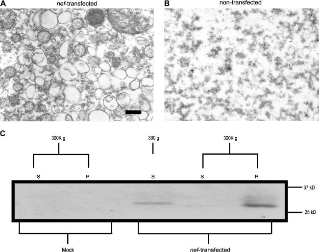
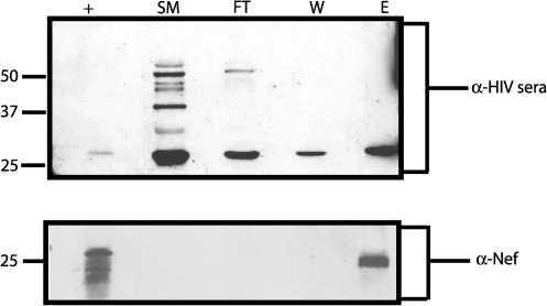
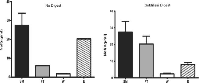
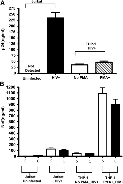
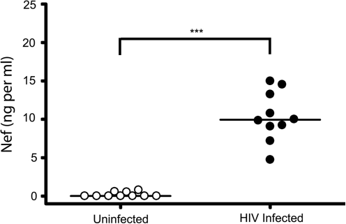
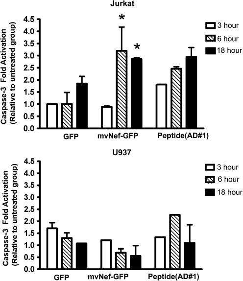
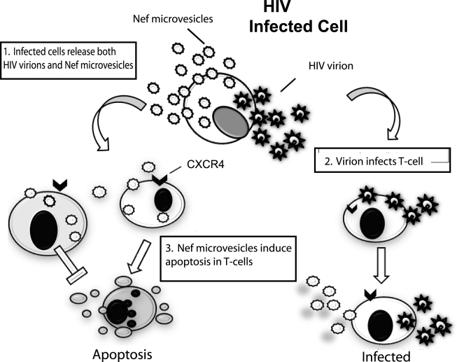
HIV-1 Nef has been demonstrated to be integral for viral persistence, infectivity, and the acceleration of disease pathogenesis (AIDS) in humans. Nef has also been detected in the plasma of HIV-infected individuals and is released from infected cells. The form in which Nef is released from infected cells is unknown. However, Nef is a myristoylated protein and has been shown to interact with the intracellular vesicular trafficking network. Here we show that Nef is released in CD45-containing microvesicles. This microvesicular Nef (mvNef) is detected in the plasma of HIV-infected individuals at relatively high concentrations (10 ng/ml). It is also present in tissue culture supernatants of Jurkat cells infected with HIV(MN). Interestingly, plasma mvNef levels in HIV(+) patients did not significantly correlate with viral load or CD4 count. Microvesicular Nef levels persisted in the plasma of HIV-infected individuals despite the use of antiretroviral therapy, even in individuals with undetectable viral loads. Using cell lines, we found Nef microvesicles induce apoptosis in Jurkat T-lymphocytes but had no observed effect on the U937 monocytic cell line. Given the large amount of mvNef present in the plasma of HIV-infected individuals, the apoptotic effect of mvNef on T cells, and the observed functions of extracellular soluble Nef in vitro, it seems likely that in vivo mvNef may play a significant role in the pathogenesis of AIDS.


Figures

FIG. 1.
FIG. 1.
Nef expression and HIV-1 infection induce vesicle formation. TEM image of conditioned media from HEK293 cells transfected with (A) Nef expression plasmid or (B) mock-transfected. Conditioned media collected 3 days posttransfection and subjected to ultracentrifugation (400,000 × g). Pellets were fixed, embedded, and stained with 2% uranyl acetate. Samples were visualized using a JEOL 1200 EX transmission microscope. (C) Culture supernatant from HEK293 transfected with a Nef expression plasmid was subjected high-speed differential centrifugation. Supernatants were centrifuged for 10 min at 300 × g, followed by 30 min at 10,000 × g, and then centrifuged for 1 h at 300,000 × g. Nef was detected in vesicles pelleted at 300,000 by immunoblot using anti-Nef monoclonal. S, supernatants; P, pellet.
FIG. 2.
FIG. 2.
Nef vesicles captured using anti-CD45 magnetic bead separation. Supernatants from HIV-1-infected cells were harvested 15 days postinfection and vesicles/virions were concentrated by centrifugation at 200,000 × g for 1 h on a 20% sucrose cushion. The pellet was resuspended in 1 × TNE buffer and subjected to CD45 magnetic bead separation. Each fraction was collected, ultracentrifuged at 400,000 × g for 1 h, and then separated by SDS-PAGE. Fractions (SM = starting material, FT = flow through, W = wash, and E = eluate) were analyzed by Western blot for HIV proteins using anti-HIV sera (upper panel). The singular band present in the eluate was confirmed to be Nef via an immunoblot using an anti-Nef monoclonal antibody (lower panel). Nef+, recombinant Nef protein.
FIG. 3.
FIG. 3.
Subtilisin treatment reduces CD45 capture of Nef microvesicles capture. Nef microvesicles from supernatants of HIV-infected Jurkat cells (15 days postinfection) were isolated as described in Fig. 2, treated for 4 h with subtilisin (2 mg/ml), and then CD45 affinity purified. Nef contents in subsequent SM, FT, W, and E fractions were measured by ELISA. SM = starting material, FT = flow through, W = wash, and E = eluate.
FIG. 4.
FIG. 4.
Production of CD45+ Nef microvesicles is cell-type dependent. Concentrations of p24 (A) microvesicles and Nef (B) were measured by ELISA in culture supernatants and CD45 captured microvesicles of HIV-1-infected Jurkat T-lymphocytes, THP-1 monocytes, and PMA-treated THP-1 (macrophage-like). T-lymphocytes release the most HIV-1 virus as measured by p24 ELISA whereas macrophage-like THP-1 releases the most Nef microvesicles.
FIG. 5.
FIG. 5.
Nef microvesicles are detected in plasma of HIV-infected subjects. Plasma was collected from uninfected (n = 15) and HIV+-infected individuals (n = 10). Microvesicles were ultracentrifuged (400,000 × g); pellets were resuspended in PBS and assayed for Nef using a Nef-capture ELISA. The median value of Nef in plasma is 10.3 (ng/1 ml plasma). Statistical significance was determined using a two-tailed Mann–Whitney analysis. ***p-value < 0.001.
FIG. 6.
FIG. 6.
Nef microvesicles in plasma of HIV-infected subjects are isolated by CD45 affinity. CD45+ microvesicles in plasma of (A) uninfected and (B) HIV+ donors were isolated as described previously in Fig. 2. Fractions generated during CD45 affinity (SM, FT, W, and E) were ultracentrifuged (400,000 × g); the pellets were resuspended in PBS and assayed for Nef via Nef capture ELISA. Nef was detected in the SM and CD45 captured material (E). SM = starting material, FT = flow through, W = wash, and E = CD45 captured eluate.
FIG. 7.
FIG. 7.
Exposure to vesicular Nef induces apoptosis. Caspase-3 activation in Jurkat and U937 monocytic cells exposed to Nef for 3, 6, and 18 h was measured using a 3-plex Luminex kit. Statistical significance was determined using ANOVA, *p-value <0.05.
FIG. 8.
FIG. 8.
Model of Nef microvesicle in HIV pathogenesis. HIV-infected cells release both virions and Nef microvesicles. Depending on the cell type of the microvesicle, the virion ratio varies. In step 1 the infected cells release both virions and Nef microvesicles. Cells (both T cells and monocytes) that come in contact with virions become infected (step 2). T cells encountering only Nef microvesicles undergo apoptosis whereas monocytes/macrophages do not (step 3). In this model Nef microvesicles are integral to HIV pathogenesis.

References

    1. Deacon NJ. Tsykin A. Solomon A, et al. Genomic structure of an attenuated quasi species of HIV-1 from a blood transfusion donor and recipients. Science. 1995;270(5238):988–991. - PubMed
    1. Kirchhoff F. Greenough TC. Brettler DB. Sullivan JL. Desrosiers RC. Brief report: Absence of intact nef sequences in a long-term survivor with nonprogressive HIV-1 infection. N Engl J Med. 1995;332(4):228–232. - PubMed
    1. Learmont JC. Geczy AF. Mills J, et al. Immunologic and virologic status after 14 to 18 years of infection with an attenuated strain of HIV-1. A report from the Sydney Blood Bank Cohort. N Engl J Med. 1999;340(22):1715–1722. - PubMed
    1. Lewin SR. Lambert P. Deacon NJ. Mills J. Crowe SM. Constitutive expression of p50 homodimer in freshly isolated human monocytes decreases with in vitro and in vivo differentiation: A possible mechanism influencing human immunodeficiency virus replication in monocytes and mature macrophages. J Virol. 1997;71(3):2114–2119. - PMC - PubMed
    1. Salvi R. Garbuglia AR. Di CA, et al. Grossly defective nef gene sequences in a human immunodeficiency virus type 1-seropositive long-term nonprogressor. J Virol. 1998;72(5):3646–3657. - PMC - PubMed

Publication types

Substances