Review
. 2011 Jan;163(1):1-10.
doi: 10.1111/j.1365-2249.2010.04261.x. Epub 2010 Oct 21.

Rapid detection, enrichment and propagation of specific T cell subsets based on cytokine secretion

Affiliations
Review

Rapid detection, enrichment and propagation of specific T cell subsets based on cytokine secretion

J D M Campbell et al. Clin Exp Immunol. 2011 Jan.

Abstract

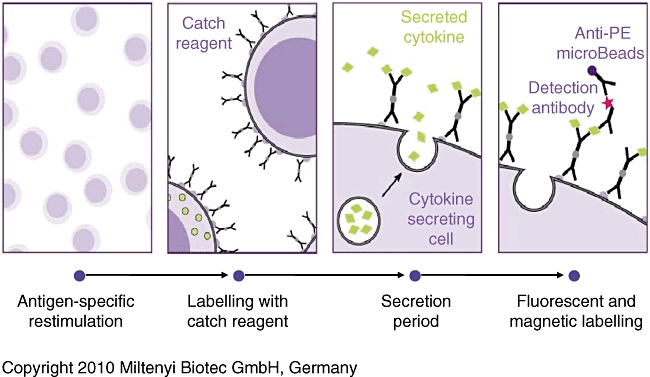
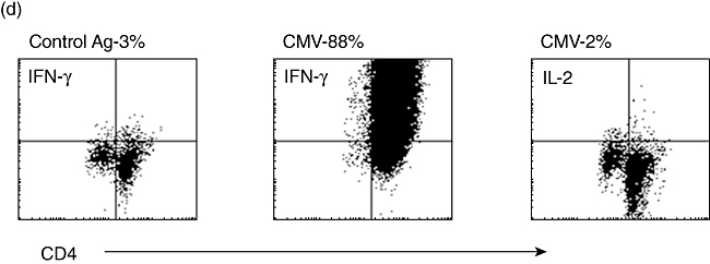
T cell lines with defined cytokine profiles are an invaluable tool for assessing the control of immune responses both in vitro and in vivo. Production of such cell lines can be complex and time-consuming. Here we present a powerful technique to assay the cytokines produced by T cells activated polyclonally or with specific antigens. This paper presents a detailed methodology for the identification and isolation of cytokine-producing T cells activated with the artificial superantigen, CytoStim, or viral and fungal antigens. These cells can be analysed for different cytokines simultaneously, or cultured further to rapidly establish T cell lines making known cytokine types. We highlight the enumeration, isolation and phenotype of interleukin-17-producing T cells, and the rapid generation of virus-specific Th1 T cell lines.


Figures

Fig. 1
Fig. 1
Summary of principal steps to label and isolate T cells using the cytokine secretion assay system.
Fig. 2
Fig. 2
Human peripheral blood mononuclear cells (PBMC) stimulated with Cytostim for 3 h or unstimulated controls, assayed with the interleukin (IL)-17 secretion assay. (a) Stimulated and unstimulated cells pre- and post-isolation over two MS columns on a mini-MACS magnet. IL-17+ cells are >90% CD4+ CD154+. (b). Cells stained with CD4 and CD161 confirm that >90% of IL-17+ cells are CD161+. (c) Cells co-processed with interferon (IFN)-γ or IL-2 secretion assays show that there is a significant population of IL2+ IL-17+ cells (box) but not of IFN-γ+ IL-17+ cells. Note that IL-2 and IFN-γ are produced in significantly larger quantities than IL-17.
Fig. 3
Fig. 3
BALB/c spleen cells stimulated with phorbol myristate acetate (PMA) and ionomycin for 3–6 h, or unstimulated controls. (a) Interleukin (IL)-17 is secreted by CD4+, CD8+γ/δ T cell receptor (TCR)+ T cells and natural killer (NK) T cells at 3 h. (b) Kinetics of IL-17 secretion by CD4+ cells. The maximum percentage positive cells (•) and maximum mean fluorescence intensity (♦) are both reached within 3–4 h of stimulus. formula image
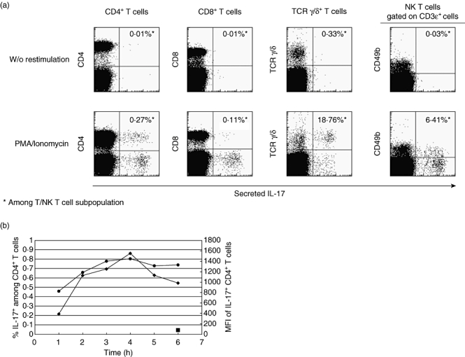
: Unstimulated controls.
Fig. 4
Fig. 4
Human peripheral blood mononuclear cells (PBMC) stimulated with Candida albicans lysate for 16 h or unstimulated controls, assayed with the interleukin (IL)-17 secretion assay. The small number of IL-17+ cells can be enriched easily more than 2000 times and are confined to the activated CD154+ cells.
Fig. 5
Fig. 5
(a) Mean [±standard error of the mean (s.e.m.)] numbers of CD4 and CD8 T cells isolated using the interferon (IFN)-γ secretion assay per 108 starting human peripheral blood mononuclear cells (PBMC) after stimulus with cytomegalovirus (CMV)-lysate for 16 h (n = 20). Addition of the human leucocyte antigen (HLA)-A2-specific NLV(495–503) peptide from pp65 increases the numbers of CD8+ cells isolated, with an apparent bystander effect on CD4+ cells (n = 8). (b) Mean (± s.e.m.) expansion rate of T cells isolated using the IFN-γ secretion assay and expanded as per Rauser et al. [9]. CD4+ cells show an advantage in expansion rates over CD8+ cells. (c) (i) HLA-A2+ human PBMC stimulated with the HLA-A2 restricted NLV(495–503) peptide from pp65 before isolation of positive cells using the IFN-γ secretion assay (day 0–0·04% of the start total of 108 PBMC – 40 000+ cells). The isolated and expanded cells after two rounds of expansion are >99% NLV(495–503) tetramer+ and have reached 108 cells in total (day 29). (ii) HLA-A2+ HLA-B7+ human PBMC stimulated with pp65 PepTivator peptide pool. Pre-stimulus B7-restricted CD8- T cells heavily outnumber A2-restricted cells (day 0). After stimulus, isolation using the IFN-γ secretion assay and one round of expansion (day 12) the very heavy bias towards the B7-restricted cells is maintained, with 34·42% of all cells B7-restricted. (d) CD4+ T cells stimulated with CMV lysate, isolated using the IFN-γ secretion assay, and expanded for 12 days post-isolation. Restimulated with autologous dendritic cells and CMV or control lysate and assayed for intracellular IL-2 or IFN-γ expression after 6 h. The vast majority make IFN-γ to CMV-restimulation but make no IL-2, confirming the effector-memory status of these helper cells.
Fig. 5
Fig. 5
(a) Mean [±standard error of the mean (s.e.m.)] numbers of CD4 and CD8 T cells isolated using the interferon (IFN)-γ secretion assay per 108 starting human peripheral blood mononuclear cells (PBMC) after stimulus with cytomegalovirus (CMV)-lysate for 16 h (n = 20). Addition of the human leucocyte antigen (HLA)-A2-specific NLV(495–503) peptide from pp65 increases the numbers of CD8+ cells isolated, with an apparent bystander effect on CD4+ cells (n = 8). (b) Mean (± s.e.m.) expansion rate of T cells isolated using the IFN-γ secretion assay and expanded as per Rauser et al. [9]. CD4+ cells show an advantage in expansion rates over CD8+ cells. (c) (i) HLA-A2+ human PBMC stimulated with the HLA-A2 restricted NLV(495–503) peptide from pp65 before isolation of positive cells using the IFN-γ secretion assay (day 0–0·04% of the start total of 108 PBMC – 40 000+ cells). The isolated and expanded cells after two rounds of expansion are >99% NLV(495–503) tetramer+ and have reached 108 cells in total (day 29). (ii) HLA-A2+ HLA-B7+ human PBMC stimulated with pp65 PepTivator peptide pool. Pre-stimulus B7-restricted CD8- T cells heavily outnumber A2-restricted cells (day 0). After stimulus, isolation using the IFN-γ secretion assay and one round of expansion (day 12) the very heavy bias towards the B7-restricted cells is maintained, with 34·42% of all cells B7-restricted. (d) CD4+ T cells stimulated with CMV lysate, isolated using the IFN-γ secretion assay, and expanded for 12 days post-isolation. Restimulated with autologous dendritic cells and CMV or control lysate and assayed for intracellular IL-2 or IFN-γ expression after 6 h. The vast majority make IFN-γ to CMV-restimulation but make no IL-2, confirming the effector-memory status of these helper cells.

References

    1. Mosmann TR, Cherwinski H, Bond MW, et al. Two types of murine helper T cell clone. I. Definition according to profiles of lymphokine activities and secreted proteins. J Immunol. 1986;136:2348–57. - PubMed
    1. Wan YY. Multi-tasking of helper T cells. Immunology. 2010;130:166–71. - PMC - PubMed
    1. O'Connor RA, Taams LS, Anderton SM. Translational mini-review series on Th17 cells: CD4 T helper cells: functional plasticity and differential sensitivity to regulatory T cell-mediated regulation. Clin Exp Immunol. 2010;159:137–47. - PMC - PubMed
    1. O'Connor RA, Prendergast CT, Sabatos CA, et al. Th1 cells facilitate the entry of Th17 cells to the central nervous system during experimental autoimmune encephalomyelitis. J Immunol. 2008;181:3750–54. - PMC - PubMed
    1. Peggs KS, Verfuerth S, Pizzey A, et al. Cytomegalovirus-specific T cell immunotherapy promotes restoration of durable functional antiviral immunity following allogeneic stem cell transplantation. Clin Infect Dis. 2009;49:1851–60. - PubMed

MeSH terms