. 2010 Nov;32(10):1618-31.
doi: 10.1111/j.1460-9568.2010.07447.x. Epub 2010 Oct 21.

Polarized targeting of L1-CAM regulates axonal and dendritic bundling in vitro

Affiliations

Polarized targeting of L1-CAM regulates axonal and dendritic bundling in vitro

Joshua Barry et al. Eur J Neurosci. 2010 Nov.

Abstract

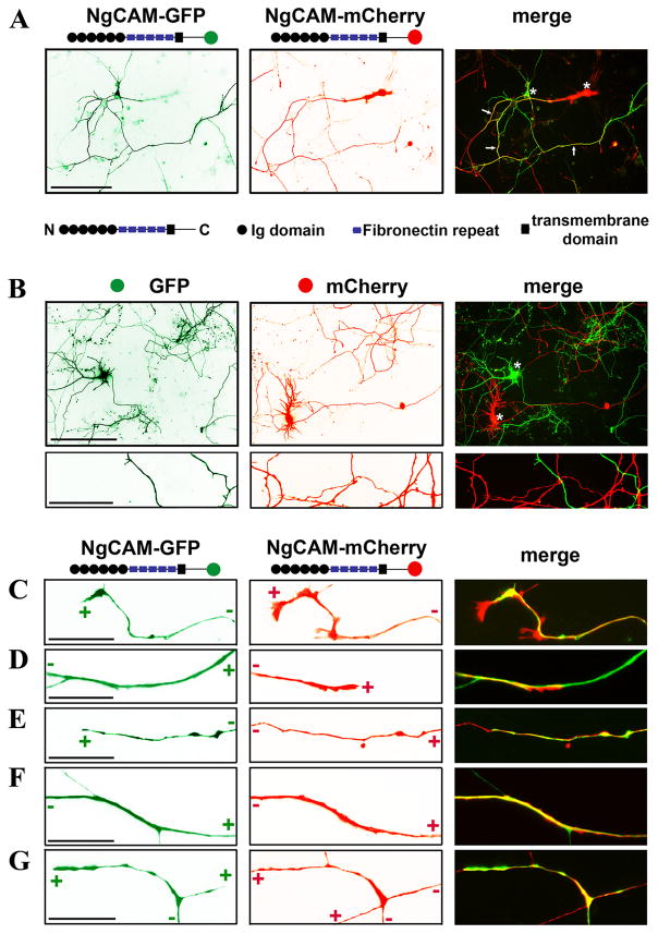
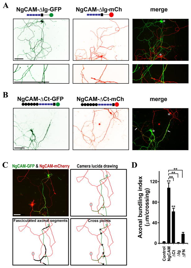
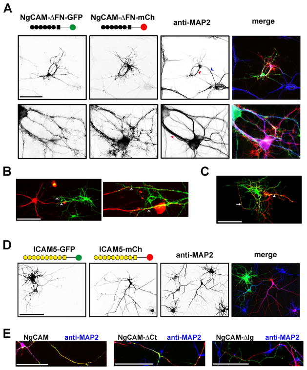
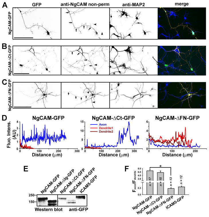
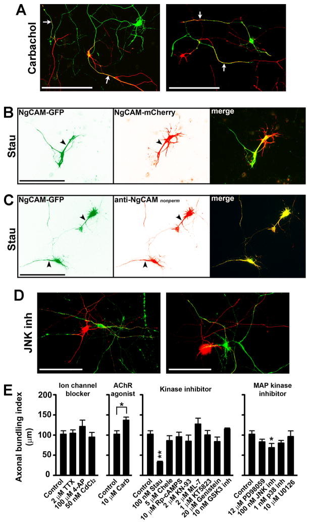
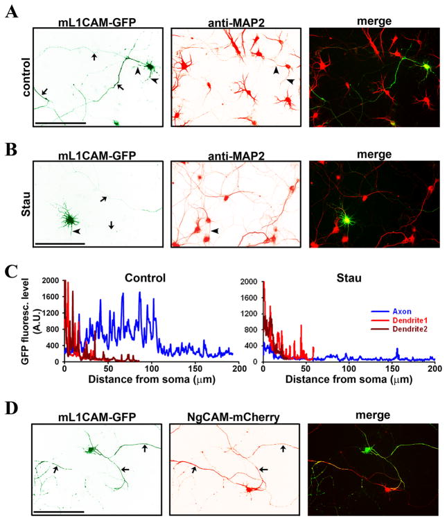
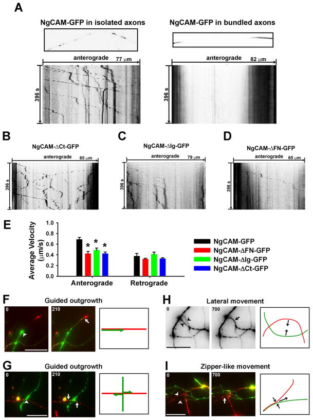
Proper axonal and dendritic bundling is essential for the establishment of neuronal connections and the synchronization of synaptic inputs, respectively. Cell adhesion molecules of the L1-CAM (L1-cell adhesion molecule) family regulate axon guidance and fasciculation, neuron migration, dendrite morphology, and synaptic plasticity. It remains unclear how these molecules play so many different roles. Here we show that polarized axon-dendrite targeting of an avian L1-CAM protein, NgCAM (neuron-glia cell adhesion molecule), can regulate the switch of bundling of the two major compartments of rat hippocampal neurons. Using a new in-vitro model for studying neurite-neurite interactions, we found that expressed axonal NgCAM induced robust axonal bundling via the trans-homophilic interaction of immunoglobulin domains. Interestingly, dendritic bundling was induced by the dendritic targeting of NgCAM, caused by either deleting its fibronectin repeats or blocking activities of protein kinases. Consistent with the NgCAM results, expression of mouse L1-CAM also induced axonal bundling and blocking kinase activities disrupted its axonal targeting. Furthermore, the trans-homophilic interaction stabilized the bundle formation, probably through recruiting NgCAM proteins to contact sites and promoting guided axon outgrowth. Taken together, our results suggest that precise localization of L1-CAM is important for establishing proper cell-cell contacts in neural circuits.


Figures

Fig. 1
Fig. 1. Expression of NgCAM fusion proteins induced robust axonal bundling in cultured hippocampal neurons
Fluorescence proteins, GFP and mCherry, were fused to the C-terminus of NgCAM. Using a modified procedure, NgCAM-GFP and NgCAM-mCherry were transfected into different neurons in culture at 5 DIV and fixed 3 days later. (A) Extensive bundling was induced between the axons of NgCAM-GFP-and NgCAM-mCherry-expressing neurons. Structural diagrams of NgCAM constructs are provided. Black circles, the Ig domains; Purple rectangles, fibronectin repeats; The black squares, the membrane-spanning segment; Green and red circles, GFP and mCherry; “N” and “C”, the extracellular N- and intracellular C-terminus. Arrows, bundled axonal segments. (B) Despite many crossings, little bundling occurred between the axons of GFP- and mCherry-expressing neurons. The images in (C)-(G) in higher magnification show representative modes of axonal bundling in our culture. (C) Co-localization of two axonal growth cones expressing NgCAM-GFP and NgCAM-mCherry. (D) An axonal growth cone grew along a pioneer axon. (E) An axonal growth cone migrated in the reverse direction along an existing axon. (F) Bundled and isolated segments of a thin axon with a thick axonal bundle. (G) Branching of bundled axons. “+” and “−” indicate the directions of axonal growth cone and soma, respectively. Asterisks, soma of transfected neurons. Scale bars, 100 μm in (A) and upper panels of (B), 10 μm in (C)-(G) and lower panels of (B).
Fig. 2
Fig. 2. Distinct roles of Ig and C-terminal domains of NgCAM in axonal bundling
(A) Deleting the Ig domains eliminated NgCAM-induced axonal bundling. Almost no axonal bundling was observed between NgCAM-ΔIg-GFP- and NgCAM-ΔIg-mCh-expressing neurons. NgCAM-ΔIg-GFP (green) and NgCAM-ΔIg-mCh (red) were transfected into different neurons at 5DIV and fixed three days later. (B) Deleting the intracellular C-terminal domain of NgCAM (NgCAM-ΔCt) moderately reduced axonal bundling. Axonal bundles were still abundantly present in culture. (C) Diagram for calculating the axonal bundling index (ABI). A pair of neurons expressing NgCAM-GFP (green) and NgCAM-mCherry (red) had bundled axonal segments. The camera lucida drawing is at the upper-right panel. Segments of axonal bundles are shown with thick black lines (lower left) and the crossings between green and red axons are indicated with asterisks (lower right). The ABI (104μm/crossing) of this pair of neurons equals to the total length of axonal bundles (624 μm) divided by the total number of crossings (6). (D) Summary of ABIs of NgCAM truncations. Control, between axons of GFP- and mCherry-expressing neurons; NgCAM, between axons of NgCAM-GFP- and NgCAM-mCherry-expressing neurons;ΔCt, between axons of NgCAM-ΔCt-GFP- and NgCAM-ΔCt-mCh-expressing neurons; ΔIg, between axons of NgCAM-ΔIg-GFP- and NgCAM-ΔIg-mCh-expressing neurons; ΔFN, between axons of NgCAM-ΔFN-GFP and NgCAM-ΔFN-mCh-expressing neurons. Asterisks, soma of transfected neurons. Scale bars, 100 μm. One-way ANOVA followed by Dunnett’s test was used for the comparison to the control group; ** P < 0.01. Additional One-way ANOVA followed by Dunnett’s test was performed for comparing the truncations to the wild type NgCAM shown on the top; ** P < 0.01.
Fig. 3
Fig. 3. Deleting the fibronectin repeats decreased axonal bundling but induced dendritic bundling
(A) The dendrites of a pair of neurons expressing NgCAM-ΔFN-GFP (green) and NgCAM-ΔFN-mCh (red) formed bundles in both parallel (blue arrowhead) and anti-parallel (red arrowhead) manners. The anti-MAP2 (microtubule associate protein 2) staining (blue in merged) indicated the dendrites and dendritic bundles. (B) Anti-parallel dendritic bundling. Dendrites of a pair of neurons expressing NgCAM-ΔFN-GFP (green) and NgCAM-ΔFN-mCh (red) grew towards each other and formed two bundles, indicated by white arrowheads. A high magnification image is provided on the right. (C) Parallel dendritic bundling. A pair of transfected neurons close to each other had an axonal bundle indicated by a white arrow and a parallel dendritic bundle indicated by a white arrowhead. (D) ICAM5 (telencephalin) constructs (ICAM5-GFP and ICAM5-mCh) were mainly localized in dendrites but did not induce dendritic bundling. Yellow circles, Ig domains; Yellow square, the membrane-spanning segment. (E) The axon-dendrite localization of NgCAM constructs revealed by dendritic marker MAP2. Neurons transfected with GFP (green) or mCherry (red) fused NgCAM constructs were stained with an anti-MAP2 antibody (blue). Scale bars, 100 μm.
Fig. 4
Fig. 4. Axon-dendrite targeting of NgCAM-GFP and its deletion mutants
Neurons transfected with NgCAM-GFP or its truncations were stained with anti-NgCAM antibody under non-permeabilized conditions, and then stained for MAP2, a dendritic marker, under permeabilized conditions. Signals are inverted in unmerged images. In merged images (right), GFP fluorescence is in green, anti-NgCAM staining in red, and anti-MAP2 staining in blue. (A) NgCAM-GFP was mainly localized on axonal membranes. (B) NgCAM-ΔCt-GFP was mainly localized on axonal membranes similar to the wildtype. (C) Deletion of the fibronectin repeat domain resulted in dendritic targeting of NgCAM-ΔFN-GFP. NgCAM-ΔFN-GFP was mainly enriched on dendritic membranes. Arrows, axons. Arrowheads, dendrites. Scale bars, 100 μm. (D) Surface levels of the three NgCAM constructs along the axon (blue) and two main dendrites (red and dark red). (E) Western blots of GFP fusion constructs expressed in HEK293 cells. Mouse monoclonal anti-GFP antibody was used. (F) Summary of polarized targeting of NgCAM and ICAM5 constructs. For NgCAM constructs the surface levels were measured, but for ICAM5-GFP the GFP fluorescence intensity reflecting the total protein level was measured. One-way ANOVA followed by Dunnett’s test was used for the comparison among three NgCAM constructs; * P < 0.001.
Fig. 5
Fig. 5. Protein phosphorylation regulated axonal targeting of NgCAM and hence neurite bundling
Hippocampal neurons transfected with either NgCAM-GFP or NgCAM-mCherry at 5 DIV were incubated with various drugs for 2 to 3 days, and then fixed and stained for quantification of polarized targeting and axonal bundling. (A) The carbachol treatment enhanced axonal bundling. White arrows, bundled axons. (B) Inhibiting protein kinase activities by the Stau treatment markedly decreased axonal bundling but increased dendritic bundling. Black arrowheads, dendrites. (C) The Stau treatment resulted in dendritic targeting of NgCAM. (D) Inhibiting the JNK kinase activity significantly decreased axonal bundling. NgCAM-GFP in green and NgCAM-mCherry in red. Scale bars, 100 μm. (E) Summary of the effects of different drugs on NgCAM-induced axonal bundling. Four panels of drug treatment experiments included the effects of ion channel blockers, AChR agonist, kinase inhibitors, and MAP kinase inhibitors, from left to right. The same control group was used in the four panels. One-way ANOVA followed by Dunnett’s test for comparing three or more groups, * P < 0.05. **P < 0.01. Two-tailed Student’s t-test for comparing two groups, * P < 0.05.
Fig. 6
Fig. 6. Mouse L1-CAM (mL1CAM) axonal targeting is regulated by protein phosphorylation and induces axonal bundling
GFP was fused to the C-terminus of mL1CAM (mL1CAM-GFP). (A) When expressed in cultured hippocampal neurons, mL1CAM-GFP (green) mainly localized in axons. Dendrites were revealed with the MAP2 staining (red). (B) After treated with Stau, mL1CAM-GFP was mainly localized in dendrites but not axons. (C) GFP fluorescence levels of control (A) and Stau-treated neurons (B) along the axon (blue) and two main dendrites (red and dark red). Background value, 230, was subtracted. (D) mL1CAM-GFP-expressing axons formed bundles with NgCAM-mCherry-expressing axons. Arrows, axons. Arrowheads, dendrites. Scale bars, 100 μm.
Fig. 7
Fig. 7. Live cell imaging of NgCAM transport and neurite movements in bundle formation
(A) NgCAM-GFP-containing carriers moving anterogradely in an isolated axon (left) and reduced axonal transport in bundled axons (right). Many NgCAM-GFP molecules reflected by the strong GFP intensity were present in vesicular- and tubular-shaped carriers. The FRAP imaging experiments are shown by one frame of the axonal segment (upper) and the kymograph (lower). In kymographs, the total time (second) is indicated on the left, the total length (μm) on the top. (B) A kymograph of NgCAM-ΔCt-GFP axonal transport. (C) A kymograph of NgCAM-ΔIg-GFP axonal transport. (D) A kymograph of NgCAM-ΔFN-GFP axonal transport. (E) Reduced average velocity of anterograde but not retrograde axonal transport of the NgCAM mutations. One-way ANOVA followed by Dunnett’s test. * p < 0.05. Long-term live-cell imaging revealed both guided axonal outgrowth ((F) and (G)) and lateral movements of axon shafts ((H) and (I)). (F) An axon (green) grew along a leading axon (red). (G) Two branches (green) were generated and grew along an existing axon (red) at a crossing. (H) Two axons moved towards each other laterally and formed a bundle. (I) Formation of a network of bundled axons included a zipper-like movement and guided axon outgrowth. Arrowheads indicate another kind of lateral movement, zipper-like movement. The first and last frames are provided, and the model diagram drawing is on the right. Arrowheads, beginning positions; Arrows, ending positions. Numbers at the upper-left corner, time in minutes. Scale bars, 20 μm.

References

    1. Bak M, Fraser SE. Axon fasciculation and differences in midline kinetics between pioneer and follower axons within commissural fascicles. Development. 2003;130:4999–5008. - PubMed
    1. Bastiani MJ, Raper JA, Goodman CS. Pathfinding by neuronal growth cones in grasshopper embryos. III. Selective affinity of the G growth cone for the P cells within the A/P fascicle. J Neurosci. 1984;4:2311–2328. - PMC - PubMed
    1. Bennett V, Chen L. Ankyrins and cellular targeting of diverse membrane proteins to physiological sites. Curr Opin Cell Biol. 2001;13:61–67. - PubMed
    1. Campbell RE, Gaidamaka G, Han SK, Herbison AE. Dendro-dendritic bundling and shared synapses between gonadotropin-releasing hormone neurons. Proc Natl Acad Sci U S A. 2009;106:10835–10840. - PMC - PubMed
    1. Cohen NR, Taylor JS, Scott LB, Guillery RW, Soriano P, Furley AJ. Errors in corticospinal axon guidance in mice lacking the neural cell adhesion molecule L1. Curr Biol. 1998;8:26–33. - PubMed

Publication types

MeSH terms