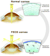
Fuchs endothelial corneal dystrophy
- PMID: 20964980
- PMCID: PMC3061348
- DOI: 10.1016/s1542-0124(12)70232-x
Fuchs endothelial corneal dystrophy
Abstract
Fuchs endothelial corneal dystrophy (FECD) is characterized by progressive loss of corneal endothelial cells, thickening of Descement's membrane and deposition of extracellular matrix in the form of guttae. When the number of endothelial cells becomes critically low, the cornea swells and causes loss of vision. The clinical course of FECD usually spans 10-20 years. Corneal transplantation is currently the only modality used to restore vision. Over the last several decades genetic studies have detected several genes, as well as areas of chromosomal loci associated with the disease. Proteomic studies have given rise to several hypotheses regarding the pathogenesis of FECD. This review expands upon the recent findings from proteomic and genetic studies and builds upon recent advances in understanding the causes of this common corneal disorder.
Conflict of interest statement
The authors have no proprietary or commercial interests in any concept or product discussed in this article.
Figures


References
-
- Fuchs E. Dystrophia epithelialis corneae. Albrecht Von Graefes Arch Klin Exp Ophthalmol. 1910;76:478–508.
-
- Iwamoto T, DeVoe AG. Electron microscopic studies on Fuchs’combined dystrophy. I. Posterior portion of the cornea. Invest Ophthalmol. 1971;10:9–28. - PubMed
-
- Waring GO, 3rd, Rodrigues MM, Laibson PR. Corneal dystrophies. II. Endothelial dystrophies. Surv Ophthalmol. 1978;23:147–168. - PubMed
-
- Waring GO, Rodrigues MM, Laibson PR. Corneal dystrophies. In: Leibowitz, editor. Corneal Disorders: Clinical Diagnosis and Management. Philadelphia, PA: Saunders; 1984.
-
- Weisenthal RW, Streeten BW. Posterior membrane dystrophies. In: Krachmer JH, Mannis MH, Holland EJ, editors. Cornea Volume II: Cornea and External Disease, Clinical Diagnosis and Management. St Louis, MO: Mosby-Year Book; 1997.
Publication types
MeSH terms
Grants and funding
LinkOut - more resources
Full Text Sources
Other Literature Sources

