. 2011 Feb 1;54(3):1887-95.
doi: 10.1016/j.neuroimage.2010.10.027. Epub 2010 Oct 18.

β-Amyloid affects frontal and posterior brain networks in normal aging

Affiliations

β-Amyloid affects frontal and posterior brain networks in normal aging

Hwamee Oh et al. Neuroimage. .

Abstract

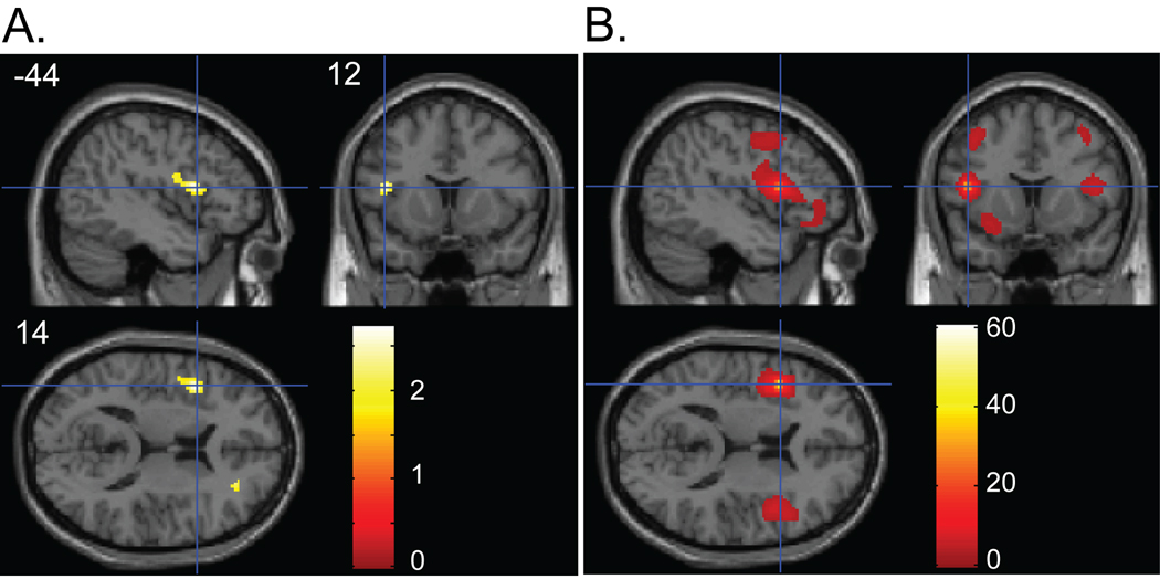
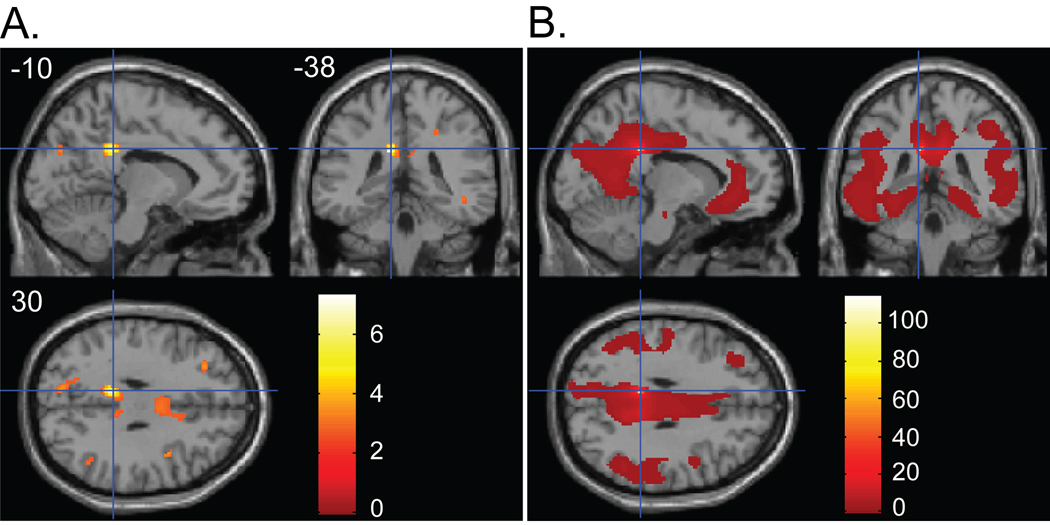
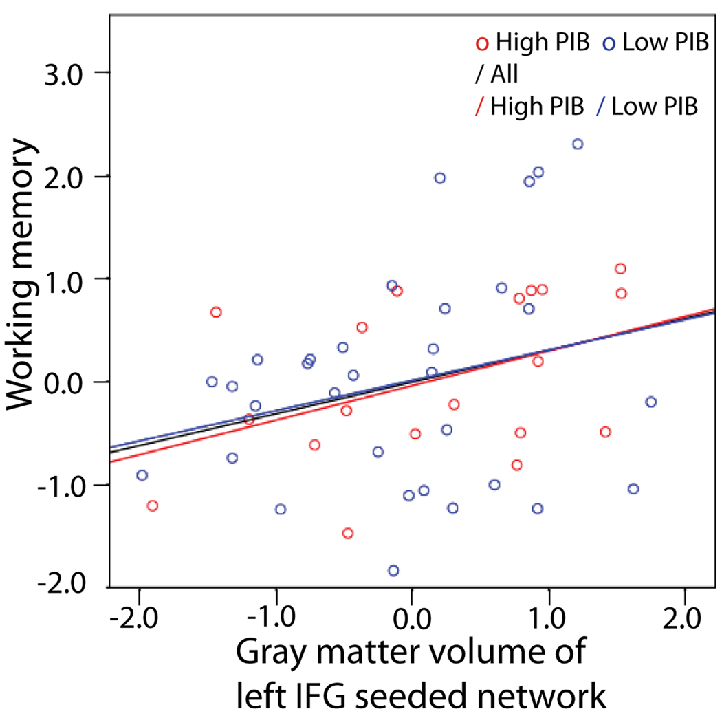
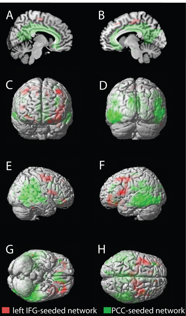
Although deposition of β-amyloid (Aβ), a pathological hallmark of Alzheimer's disease (AD), has also been reported in cognitively intact older people, its influence on brain structure and cognition during normal aging remains controversial. Using PET imaging with the radiotracer Pittsburgh compound B (PIB), structural MRI, and cognitive measures, we examined the relationships between Aβ deposition, gray matter volume, and cognition in older people without AD. Fifty-two healthy older participants underwent PIB-PET and structural MRI scanning and detailed neuropsychological tests. Results from the whole-brain voxel-based morphometry (VBM) analysis revealed that gray matter volume in the left inferior frontal cortex was negatively associated with amyloid deposition across all participants whereas reduced gray matter volume was shown in the posterior cingulate among older people with high amyloid deposition. When gray matter density measures extracted from these two regions were related to other brain regions by applying a structural covariance analysis, distinctive frontal and posterior brain networks were seen. Gray matter volume in these networks in relation to cognition, however, differed such that reduced frontal network gray matter volume was associated with poorer working memory performance while no relationship was found for the posterior network. The present findings highlight structural and cognitive changes in association with the level of Aβ deposition in cognitively intact normal elderly and suggest a differential role of Aβ-dependent gray matter loss in the frontal and posterior networks in cognition during normal aging.


Figures

Figure 1
Figure 1
(A) Regions showing a negative association between gray matter volume and PIB index. Panel shows sagittal, coronal and axial slices in MNI space with a cross hair marking the location of the average peak coordinate as indicated by numbers (X = −44, Y = 12, Z = 14) in mm. Age, gender, and TIV were controlled for. Threshold at p < .05 (uncorrected) is used for visualization. (B) Structural covariance pattern that fluctuates with gray matter density in the left IFG. Panel shows sagittal, coronal and axial slices with a cross hair marking the location of the seed region (i.e., left IFG). Age, gender, and TIV were controlled for. The map was thresholded at p < .01 with multiple comparison correction (FDR). The color bars represent T-values.
Figure 2
Figure 2
(A) Regions showing a negative association between gray matter volume and PIB index in High PIB group. Panel shows sagittal, coronal and axial slices in MNI space with a cross hair marking the location of the average peak coordinate as indicated by numbers (X = −10, Y = −38, Z = 30) in mm. Age, gender, and TIV were controlled for. Threshold at p<.01(uncorrected) is used for visualization. (B) Structural covariance pattern that fluctuates with gray matter density in the PCC. Panel shows sagittal, coronal and axial slices with a cross hair marking the location of the seed region (i.e., PCC). Age, gender, and TIV were controlled for. The map was thresholded at p<.01 with multiple comparison correction (FDR). The color bars represent T-values.
Figure 3
Figure 3
A partial correlation scatterplot illustrating the relationship of frontal network grey matter volume and working memory performance for the whole group, high PIB group, and low PIB group. The x-axis of the scatterplot represents standardized residuals of frontal network grey matter volume and the y-axis represents standardized residuals of working memory factor scores. For both standardized residual measures, age, gender, education, and TIV were regressed out. The trend of the relationship between frontal gray matter volume and working memory performance is the same across all participants regardless of their PIB group membership. High PIB group: R = .432, p = .065; Low PIB group: R = .257, p = .156; Whole group: R = .308, p = .028.
Figure 4
Figure 4
Dissociation of the left IFG – seeded network and the PCC – seeded network. Group composite maps are shown on the medial view of right and left hemispheres (A and B), the anterior and posterior surfaces (C and D), the right and left lateral surfaces (E and F), and the ventral and dorsal surfaces (G and H) of the rendered MNI single-subject brain. Regions in red and green indicate suprathreshold voxels identified by structural covariance analysis with the left IFG seed (“left IFG - seeded network”) and the PCC seed (“PCC – seeded network”), respectively. Maps were thresholded at p < .01, corrected for multiple comparisons (voxel-level FDR).

References

    1. Agosta F, Vossel KA, Miller BL, Migliaccio R, Bonasera SJ, Filippi M, et al. Apolipoprotein E epsilon4 is associated with disease-specific effects on brain atrophy in Alzheimer's disease and frontotemporal dementia. Proc Natl Acad Sci U S A. 2009;106(6):2018–2022. - PMC - PubMed
    1. Aizenstein HJ, Nebes RD, Saxton JA, Price JC, Mathis CA, Tsopelas ND, et al. Frequent amyloid deposition without significant cognitive impairment among the elderly. Arch Neurol. 2008;65(11):1509–1517. - PMC - PubMed
    1. Archer HA, Edison P, Brooks DJ, Barnes J, Frost C, Yeatman T, et al. Amyloid load and cerebral atrophy in Alzheimer's disease: an 11C-PIB positron emission tomography study. Ann Neurol. 2006;60(1):145–147. - PubMed
    1. Ashburner J, Friston KJ. Nonlinear spatial normalization using basis functions. Hum Brain Mapp. 1999;7(4):254–266. - PMC - PubMed
    1. Ashburner J, Friston KJ. Voxel-based morphometry--the methods. Neuroimage. 2000;11(6):805–821. - PubMed

MeSH terms