. 2011 Mar;232(1-2):17-25.
doi: 10.1016/j.jneuroim.2010.09.025. Epub 2010 Oct 20.

Localized retinal neuropeptide regulation of macrophage and microglial cell functionality

Affiliations

Localized retinal neuropeptide regulation of macrophage and microglial cell functionality

Norikuni Kawanaka et al. J Neuroimmunol. 2011 Mar.

Abstract

The functionality of immune cells is manipulated within the ocular microenvironment to protect the sensitive and non-regenerating light-gathering tissue from the collateral damage of inflammation. This is mediated partly by the constitutive presence of immunomodulating neuropeptides. Treating primary resting macrophages with soluble factors produced by the posterior eye induced co-expression of Arginase1 and NOS2. The neuropeptides alpha-melanocyte stimulating hormone and Neuropeptide Y alternatively activated the macrophages to co-express Arginase1 and NOS2 like myeloid suppressor cells. Similar co-expressing cells were found within healthy, but not in wounded retinas. Therefore, the healthy retina regulates macrophage functionality to the benefit of ocular immune privilege.


Figures

Figure 1
Figure 1
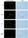
Expression of Arginase1, NOS2, and TUNEL staining in primary macrophages treated with eyecup conditioned media. Primary macrophages were treated with conditioned media (CM), CM depleted of α-MSH (−α-MSH), CM depleted of NPY (−NPY), or CM depleted of both α-MSH and NPY (−α-MSH −NPY). The treated cells were incubated for 24 hours, and then co-stained with FITC-labeled anti-Arginase1 and PE-anti-NOS2, or assayed for apoptosis by TUNEL staining methods (red stained cells). The results are representative of three separate experiments with three different conditioned media.
Figure 2
Figure 2
Expression of Arginase1 and NOS2 by macrophages treated with conditioned media of laser-wounded or POMC knock out (POMC(−/−)) eyes. The concentration of (A) α-MSH, and (B) NPY in healthy and laser-wounded CM were assayed by ELISA. Presented are the mean ± SEM of four eyecup CM from four different mice. *Statistical difference (P < 0.001) was determined by Student's T-test. NS= not significantly different. Primary macrophages were treated for 24 hours with (C, G) CM of laser-wounded RPE eyecups, or (D, H) CM of laser-wounded RPE eyecups plus 1 ng/ml of α-MSH added. The treated cells were co-stained with FITC-labeled anti-Arginase1 and PE-anti-NOS2 (C, D), or assayed by TUNEL for apoptosis (G, H). The results are representative of three separate experiments with three different conditioned media. In addition, Primary macrophages were treated with either (E, I) CM of POMC(−/−) eyecups, or (F, J) CM of POMC(−/−) eyecups plus 1 ng/ml of α-MSH added. The treated cells were co-stained with FITC-labeled anti-Arginase1 and PE-anti-NOS2 (E, F), or assayed by TUNEL for apoptosis (I, J). The results are representative of two separate experiments with two different conditioned media.
Figure 3
Figure 3
The effects of treating macrophages with α-MSH and NPY. Primary macrophages were treated with α-MSH (1ng/ml), NPY (1ng/ml), or both for 24 hours. The cells were co-stained for Arginase1 and NOS2, or assayed by TUNEL staining for apoptosis. All images were made with the same exposure time and subtraction of background. The results are representative of three separate experiments.
Figure 4
Figure 4
Cytokine pattern of neuropeptide treated macrophages. The primary macrophages were untreated, treated with both α-MSH and NPY, or with LPS. After 48 hours incubation the culture supernatants were assayed for cytokine production using multi-plex analysis. Presented are the results of four independent macrophage samples (mean ± SD). Significant differences († P<0.05 or * P < 0.001) was determined by Student's T-test analysis in comparison with the LPS-stimulated macrophages.
Figure 5
Figure 5
The co-expression of Arginase1 and NOS2 in healthy and wounded retinas. Paraffin sections of C57BL/6 mice A) healthy retinas or B) 72 hour laser-wounded retinas were co-stained with FITC-labeled anti-Arginase1 and PE-anti-NOS2. Plain arrows identify double stained cells, and the * arrows mark single stained cells. The expression of nitrotyrosine in C) healthy and D) wounded retinas were done on retinal sections co-stained for nitrotyrosine, and for the macrophage/microglial cell marker CD64. The arrows identify CD65+ cells, and * arrows identify CD65+ cells that are not associated with nitrotyrosine staining. Inserts are magnified regions boxed in the figure. The results are representative sections selected from three different sections from three different mice.
Figure 5
Figure 5
The co-expression of Arginase1 and NOS2 in healthy and wounded retinas. Paraffin sections of C57BL/6 mice A) healthy retinas or B) 72 hour laser-wounded retinas were co-stained with FITC-labeled anti-Arginase1 and PE-anti-NOS2. Plain arrows identify double stained cells, and the * arrows mark single stained cells. The expression of nitrotyrosine in C) healthy and D) wounded retinas were done on retinal sections co-stained for nitrotyrosine, and for the macrophage/microglial cell marker CD64. The arrows identify CD65+ cells, and * arrows identify CD65+ cells that are not associated with nitrotyrosine staining. Inserts are magnified regions boxed in the figure. The results are representative sections selected from three different sections from three different mice.

Similar articles

Cited by

References

    1. Alvaro AR, Rosmaninho-Salgado J, Santiago AR, Martins J, Aveleira C, Santos PF, Pereira T, Gouveia D, Carvalho AL, Grouzmann E, Ambrosio AF, Cavadas C. NPY in rat retina is present in neurons, in endothelial cells and also in microglial and Muller cells. Neurochem Int. 2007;50:757–763. - PubMed
    1. Ammar DA, Hughes BA, Thompson DA. Neuropeptide Y and the retinal pigment epithelium: receptor subtypes, signaling, and bioelectrical responses. Invest Ophthalmol Vis Sci. 1998;39:1870–1878. - PubMed
    1. Bohm M, Wolff I, Scholzen TE, Robinson SJ, Healy E, Luger TA, Schwarz T, Schwarz A. Alpha-melanocyte-stimulating hormone protects from ultraviolet radiation-induced apoptosis and DNA damage. J Biol Chem. 2004 - PubMed
    1. Bronte V, Zanovello P. Regulation of immune responses by L-arginine metabolism. Nat Rev Immunol. 2005;5:641–654. - PubMed
    1. Bruun A, Tornqvist K, Ehinger B. Neuropeptide Y (NPY) immunoreactive neurons in the retina of different species. Histochemistry. 1986;86:135–140. - PubMed

Publication types

MeSH terms