Comparative Study
. 2012 Jun;33(6):1034-44.
doi: 10.1016/j.neurobiolaging.2010.08.016. Epub 2010 Oct 20.

Elevated 4-hydroxyhexenal in Alzheimer's disease (AD) progression

Affiliations
Comparative Study

Elevated 4-hydroxyhexenal in Alzheimer's disease (AD) progression

Melissa A Bradley et al. Neurobiol Aging. 2012 Jun.

Abstract

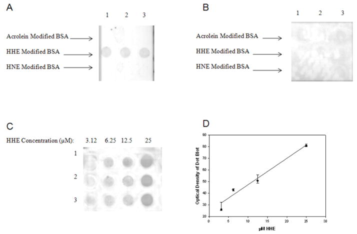
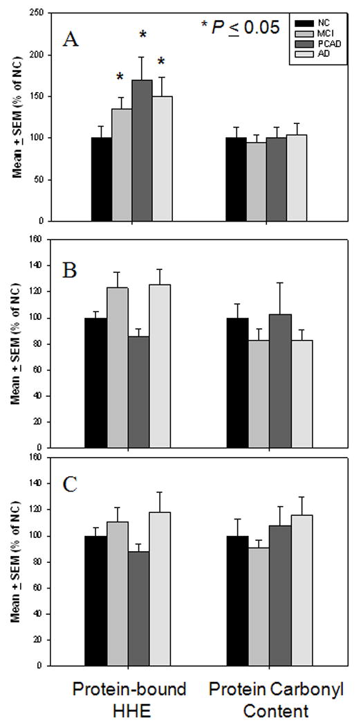
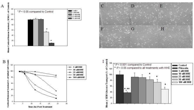
Multiple studies have demonstrated elevations of α, β-unsaturated aldehydes including 4-hydroxynonenal (HNE) and acrolein, in vulnerable regions of mild cognitive impairment (MCI), preclinical Alzheimer's disease (PCAD), and late stage Alzheimer's disease (LAD) brain. However, there has been limited study of a third member, 4-hydroxyhexenal (HHE), a diffusible lipid peroxidation product of the ω-3 polyunstataturated fatty acids (PUFAs). In the present study levels of extractable and protein-bound HHE were quantified in the hippocampus/parahippocampal gyrus (HPG), superior and middle temporal gyri (SMTG), and cerebellum (CER) of MCI, PCAD, LAD, and normal control (NC) subjects. Levels of extractable and protein-bound HHE were increased in multiple regions in the progression of Alzheimer's disease (AD). Extractable HHE was significantly elevated in the hippocampus/parahippocampal gyrus (HPG) of PCAD and LAD subjects and protein-bound HHE was significantly higher in MCI, PCAD, and LAD HPG. A time- and concentration-dependent decrease in survival and a concentration-dependent decrease in glucose uptake were observed in primary cortical cultures treated with HHE. Together these data support a role for lipid peroxidation in the progression of Alzheimer's disease.


Conflict of interest statement

Conflict of Interest The authors have no conflicts of interest.

Figures

Figure 1
Figure 1
Specificity of mouse monoclonal antibody against HHE. Acrolein modified BSA loaded in triplicate in Row 1, HHE modified BSA loaded in triplicate in Row 2, and HNE modified BSA loaded in triplicate in Row 3 incubated with anti-HHE (A) or with anti-HHE pre-incubated with HHE (B). Figure 1C shows representative dot blots of HHE modified BSA with increasing HHE concentrations (3.125, 6.25, 12.5, and 25 μM). Each modified BSA sample was loaded in triplicate. Figure 1D shows a linear response between HHE concentration and immunochemical response (r = 0.94, P < 0.05).
Figure 2
Figure 2
(A) Levels of extractable HHE expressed as nmol/mg of protein in HPG of NC, MCI, PCAD, and LAD subjects. There was a significant (P ≤ 0.05) increase in the level of extractable HHE in HPG of PCAD and LAD subjects compared to NC subjects. No significant difference was observed in the level of extractable HHE in HPG of MCI subjects compared to NC subjects. (B) Levels of extractable HHE expressed as nmol/mg of protein in SMTG of NC, MCI, PCAD, and LAD subjects. There was no significant difference in levels of extractable HHE in SMTG of MCI, PCAD, or AD subjects compared to NC subjects. (C) Levels of extractable HHE expressed as nmol/mg protein in CER of NC, MCI, PCAD, and LAD subjects. There was a significant (P > 0.01) elevation in CER of LAD subjects compared to NC, MCI, and PCAD subjects. No significant changes were observed in the level of extractable HHE in CER of MCI or PCAD subjects compared to NC subjects.
Figure 3
Figure 3
Levels of protein-bound HHE and protein carbonyl content expressed as mean ± SEM (% NC). (A) Levels of protein-bound HHE and protein carbonyl content in HPG of NC, MCI, PCAD, and LAD subjects. There was a significant (P ≤ 0.05) elevation in protein-bound HHE in HPG of MCI, PCAD, and LAD subjects compared to NC subjects. No significant changes were observed in levels protein carbonyl content in any disease in the HPG. (B) Levels of protein-bound HHE and protein carbonyl content in SMTG of NC, MCI, PCAD, and LAD subjects. No significant changes were observed in either levels protein-bound HHE or protein carbonyl content in any disease stage in the SMTG. (C) Levels of protein-bound HHE and protein carbonyl content in CER of NC, MCI, PCAD and LAD subjects. No significant changes were observed in levels of protein-bound HHE or protein carbonyl content in any disease stage in the CER.
Figure 4
Figure 4
(A) Survival of primary cortical neurons in response to treatment with 4-hydroxyhexenal (HHE) at 1 μM, 10 μM, 25 μM, and 50 μM for 16 hr by the reduction of reduction of 3-(4,5-dimethylthiazol-2-yl)-2,5-diphenyltetrazodium bromide (MTT). Survival was significantly reduced (P < 0.05) at 16 hr when treated with 25 μM and 50 μM HHE compared to control cultures. (B) Survival of primary cortical neurons in response to treatment with 4-hydroxyhexenal (HHE) as assessed by cell counts at 0, 3, 6, 12, and 24 hr in response to increasing concentrations of HHE treatment (1, 10, 25, 50, and 100 μM). A time and concentration dependent decrease in cell survival was observed with HHE treatment. Phase Contrast micrographs of primary cortical neurons at 6 hr incubation period with increasing concentration of HHE: (C) Control cultures, (D) Cultures treated with 1 μM HHE, (E) Cultures treated with 10 μM HHE, (F) Cultures treated with 25 μM HHE, (G) Cultures treated with 50 μM HHE, and (H) Cultures treated with 100 μM HHE. Absence of vacuolization of cell bodies and extensive neurite fragmentation indicates that no concentration of HHE at 6 hr incubation resulted in cell death. (H) Glucose uptake in primary cortical cultures treated with increasing concentrations of HHE (0, 1, 10, 25, 50, 100 μM of HHE) for 6 hrs or with phloretin for 1 hr. Treatment with 25, 50, and 100 μM of HHE resulted in a significant (P < 0.001) decrease in glucose uptake compare to control cultures. Uptake of glucose in cultures treated with phloretin was significantly (P < 0.05) decreased compared to control cultures and cultures treated with all concentrations of HHE.

References

    1. Bonifacino JS, et al., editors. Current Protocols in Immunology. New York: John Wiley; 2001.
    1. Braak H, Braak E, Bohl J, Lang W. Alzheimer’s disease: amyloid plaques in the cerebellum. J Neurol Sci. 1989;93:277–287. - PubMed
    1. Bradley MA, Markesbery WR, Lovell MA. Increase levels of 4-hydroxynonenal and acrolein in the brain in preclinical Alzheimer’s disease (PCAD) Free Radic Biol Med In Press 2010 - PMC - PubMed
    1. Butterfield DA, Reed T, Perluigi M, De Marco C, Coccia R, Cini C, Sultana R. Elevated protein-bound levels of the lipid peroxidation product, 4-hydroxy-2-noenal, in brain from persons with mild cognitive impairment. Neurosci Lett. 2006;397:170–173. - PubMed
    1. Calingansan NY, Uchida K, Gibson GE. Protein-bound acrolein: a novel marker of oxidative stress in Alzheimer's disease. J Neurochem. 1999;72:751–756. - PubMed

Publication types