. 2011 Jan;39(Database issue):D718-23.
doi: 10.1093/nar/gkq962. Epub 2010 Oct 21.

3did: identification and classification of domain-based interactions of known three-dimensional structure

Affiliations

3did: identification and classification of domain-based interactions of known three-dimensional structure

Amelie Stein et al. Nucleic Acids Res. 2011 Jan.

Abstract

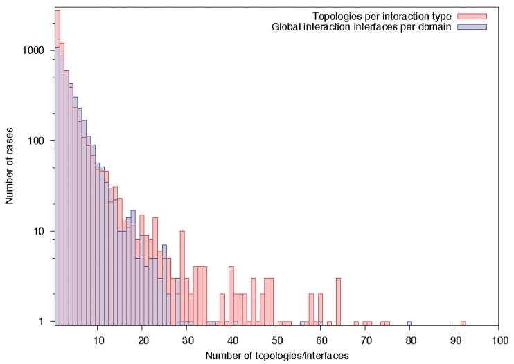
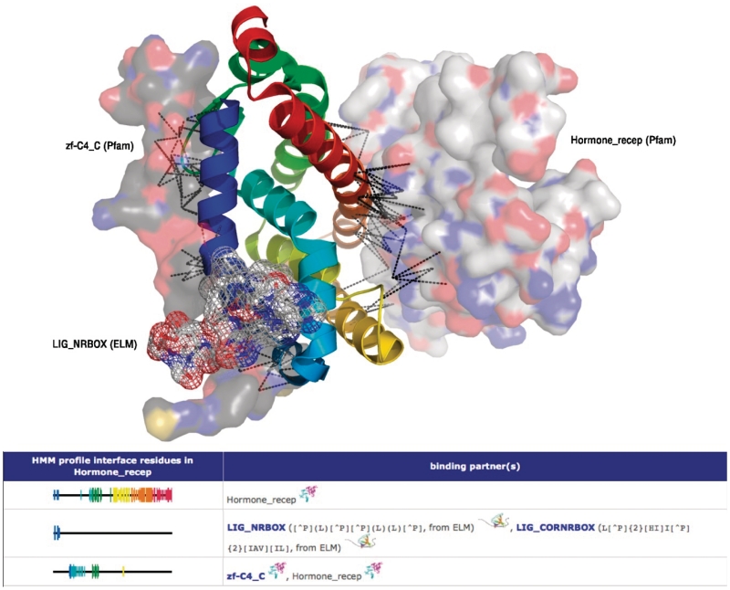
The database of three-dimensional interacting domains (3did) is a collection of protein interactions for which high-resolution three-dimensional structures are known. 3did exploits the availability of structural data to provide molecular details on interactions between two globular domains as well as novel domain-peptide interactions, derived using a recently published method from our lab. The interface residues are presented for each interaction type individually, plus global domain interfaces at which one or more partners (domains or peptides) bind. The 3did web server at http://3did.irbbarcelona.org visualizes these interfaces along with atomic details of individual interactions using Jmol. The complete contents are also available for download.


Figures

Figure 1.
Figure 1.
Flat and elongated nature of bound peptides. The chromatin assembly factor 1 peptide [magenta, PDB:1s4z (32)], bound to a chromo shadow domain in HP1beta, illustrates the typical flat and elongated structure that many peptides assume upon binding to their recognition domain. We have recently exploited this feature, together with other structural properties of peptide-mediated interactions, to identify ‘hidden’ cases of such binding events in the PDB (26).
Figure 2.
Figure 2.
Domain–domain interactions with interface topologies and structure display. For the selected domain pair, 3did displays all topologies observed in the different instances of this interaction type in all 3D structures, sorted by their frequency. Each ‘Interface Topology’ has an identifier (ID) of the form X:Y that is composed of the respective cluster IDs of the two domains. Here, the most common topology for Ras:RhoGEF is 0:2, while all other observed topologies are much less frequent. For homomeric interactions, X:X marks a symmetric topology. The ‘rainbow’ color scheme is used to indicate where interface residues lie in the sequence, from N-terminus (blue) to C-terminus (red), based on alignment to the HMM profile of the respective domains (see main text). The 3D structure of the selected instance is displayed using Jmol next to the topologies, while the interaction details (PDB ID, domain positions, score, Z-score, topology ID) are listed below. Users can select the 3D structure to be shown by clicking on the Jmol icon in the corresponding row. In domain–domain interactions, the two domains are shown in magenta and cyan, while peptide-binding domains are shown in a rainbow colour scheme to match the interface visualization.
Figure 3.
Figure 3.
Number of domain interfaces. About half of the domain–domain interactions currently stored in 3did are only observed in one interaction topology, and only a small fraction shows ten or more different binding orientations. Similarly, roughly 50% of the domains in 3did have one or two global binding interfaces, while few have 10 interfaces or more.
Figure 4.
Figure 4.
Multiple binding interfaces and their visualization. The upper part of the figure shows the hormone_recep domain (‘cartoon’ representation, rainbow colour scheme) and three of its binding partners, two domains (‘surface’ representation) and one linear motif (‘mesh’ representation). Below, the global interfaces are visualized as on the web page. These interfaces group binding partners using the same or largely overlapping interfaces, and indicates which profile positions are involved in the binding. This allows quick identification of possible overlaps, and thus competition, of different binding partners. Like the individual topologies, global interfaces are displayed in a blue-to-red rainbow colour scheme. The height of the position bars indicates how many binding partners use this particular position. By clicking on a binding partner, the user is redirected to that element’s page, while the interaction symbol (two domains or a domain and a peptide) leads to the respective interaction page (cf. Figure 2).

References

    1. Rual JF, Venkatesan K, Hao T, Hirozane-Kishikawa T, Dricot A, Li N, Berriz GF, Gibbons FD, Dreze M, Ayivi-Guedehoussou N, et al. Towards a proteome-scale map of the human protein–protein interaction network. Nature. 2005;437:1173–1178. - PubMed
    1. Stelzl U, Worm U, Lalowski M, Haenig C, Brembeck FH, Goehler H, Stroedicke M, Zenkner M, Schoenherr A, Koeppen S, et al. A human protein–protein interaction network: a resource for annotating the proteome. Cell. 2005;122:957–968. - PubMed
    1. Ewing RM, Chu P, Elisma F, Li H, Taylor P, Climie S, McBroom-Cerajewski L, Robinson MD, O'Connor L, Li M, et al. Large-scale mapping of human protein–protein interactions by mass spectrometry. Mol. Syst. Biol. 2007;3:89. - PMC - PubMed
    1. Stein A, Russell RB, Aloy P. 3did: interacting protein domains of known three-dimensional structure. Nucleic Acids Res. 2005;33:D413–D417. - PMC - PubMed
    1. Winter C, Henschel A, Kim WK, Schroeder M. SCOPPI: a structural classification of protein–protein interfaces. Nucleic Acids Res. 2006;34:D310–D314. - PMC - PubMed

Publication types