. 2010 Nov 15;24(22):2531-42.
doi: 10.1101/gad.1984210. Epub 2010 Oct 21.

Dynamic regulation of the PR-Set7 histone methyltransferase is required for normal cell cycle progression

Affiliations

Dynamic regulation of the PR-Set7 histone methyltransferase is required for normal cell cycle progression

Shumin Wu et al. Genes Dev. .

Abstract

Although the PR-Set7/Set8/KMT5a histone H4 Lys 20 monomethyltransferase (H4K20me1) plays an essential role in mammalian cell cycle progression, especially during G2/M, it remained unknown how PR-Set7 itself was regulated. In this study, we discovered the mechanisms that govern the dynamic regulation of PR-Set7 during mitosis, and that perturbation of these pathways results in defective mitotic progression. First, we found that PR-Set7 is phosphorylated at Ser 29 (S29) specifically by the cyclin-dependent kinase 1 (cdk1)/cyclinB complex, primarily from prophase through early anaphase, subsequent to global accumulation of H4K20me1. While S29 phosphorylation did not affect PR-Set7 methyltransferase activity, this event resulted in the removal of PR-Set7 from mitotic chromosomes. S29 phosphorylation also functions to stabilize PR-Set7 by directly inhibiting its interaction with the anaphase-promoting complex (APC), an E3 ubiquitin ligase. The dephosphorylation of S29 during late mitosis by the Cdc14 phosphatases was required for APC(cdh1)-mediated ubiquitination of PR-Set7 and subsequent proteolysis. This event is important for proper mitotic progression, as constitutive phosphorylation of PR-Set7 resulted in a substantial delay between metaphase and anaphase. Collectively, we elucidated the molecular mechanisms that control PR-Set7 protein levels during mitosis, and demonstrated that its orchestrated regulation is important for normal mitotic progression.


Figures

Figure 1.
Figure 1.
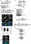
Cdk1/cyclinB phosphorylates S29 of PR-Set7. (A) Phosphorylated proteins were isolated from cells using a phosphoprotein purification column. The indicated amounts of the input, column-bound, and unbound material were fractionated by SDS-PAGE prior to Western analysis. (B) HEK-293 cells expressing Flag-PR-Set7 or S29A mutant were immunoprecipitated prior to phosphoprotein purification. Western analysis was performed using the indicated amounts of the input, column-bound, and unbound material. (C) Peptide sequences of PR-Set7 were aligned from the indicated animals using ClustalX. The conserved cdk1/cyclinB consensus sequence (S-P-X-K/R) and APC recognition motif D-box are illustrated. (D) Western analysis for PR-Set7 and pS29-PR-Set7 on lysates from cells treated with vehicle DMSO, the cdk1 inhibitor CGP74514A, or the CKI inhibitor D4476. A general H4 antibody was used to control for loading. (E) Western analyses using the indicated antibodies were performed on cells transfected with a control shRNA or two different cyclinB shRNA plasmids. (F) Recombinant wild-type PR-Set7 (WT) or N-terminal (amino acids 1–191) or C-terminal (amino acids 192–352) truncations was used as the substrate in in vitro kinase assays with purified cdk1/cyclinB. The reactions were fractionated by SDS-PAGE followed by autoradiography. (G) Western analysis of in vitro kinase assays using wild-type PR-Set7 as the substrate and either cdk1/cyclinB or CKI.
Figure 2.
Figure 2.
PR-Set7 methyltransferase activity is not altered by S29 phosphorylation. (A) HMT assays were performed using recombinant PR-Set7 or in vitro phosphorylated PR-Set7 on core histone or nucleosomal substrates and were analyzed by autoradiography or scintillation counting. Error bars represent standard deviation generated from three independent biological replicates. (B) The indicated Flag-PR-Set7 fusion proteins were immunoprecipitated prior to an HMT assay using nucleosomal substrates and were analyzed by autoradiography or scintillation counting. Error bars represent standard deviation generated from three independent biological replicates. (C) Cells treated with vehicle DMSO or the cdk1 inhibitor CGP74514A were immunostained with an H4K20me1-specific antibody (red) and counterstained with DAPI (blue).
Figure 3.
Figure 3.
Phosphorylation of S29 during prophase to anaphase results in removal of PR-Set7 from mitotic chromosomes. (A) Western analysis of cells that were chemically arrested at G1/S, released, and collected at the indicated time points and corresponding cell cycle phases. (B,C) Immunostaining using the pS29-PR-Set7 antibody (B, red) or PR-Set7 antibody (C) in the presence of vehicle DMSO or cdk1 inhibitor CGP74514A. Counterstaining with DAPI (green) was used to identify cells in interphase (a), prophase (b), metaphase (c), early anaphase (d), and telophase (e). Bar, 20 μm. (D) Flow chart of nuclear fractionation used to isolate euchromatin (S1), heterochromatin (S2), or insoluble chromatin (P). (E) Nuclei were digested with MNase for the indicated times prior to fractionation and DNA electrophoresis. The bands correspond to the expected sizes of mono-, di-, tri-, and oligonucleosomes. (F) Western analysis of the different MNase-digested fractions from cells treated with or without the cdk1 inhibitor CGP74514A.
Figure 4.
Figure 4.
APCcdh1-mediated ubiquitination and degradation of PR-Set7 is directly inhibited by S29 phosphorylation. (A) Western analysis of cells treated with CHX for the indicated time points in the presence or absence of MG132. A general H4 antibody was used as a loading control. (B) Western analysis of cells treated with CHX for the indicated time points. (C) Cells were treated with vehicle DMSO or cdk1 inhibitor CGP74514A prior to CHX treatment. Cells were collected at the indicated time points for Western analysis. (D) Cells transfected with GFP-PR-Set7 wild type, an S29A mutant, or an S29D phosphomimic mutant were counter stained with DAPI and visualized. (E) Cellular distribution of the GFP fusion proteins from D were determined in a blinded study of 200 random GFP-positive cells. (F) GFP-PR-Set7 S29A transfected cells were treated with vehicle DMSO or MG132 proteasome inhibitor and visualized. (G) The indicated Flag-PR-Set7 fusion proteins were immunoprecipitated from cells, followed by Western analysis. The asterisk indicates polyubiquitinated PR-Set7. (H) Recombinant N-terminal PR-Set7 (amino acids 1–191) was incubated ± purified APCcdh1 at the indicated time points prior to Western analysis. Increasing degrees of PR-Set7 ubiquitination are shown. (I,J) Autoradiography of 35S-labeled wild-type PR-Set7 (I) or the S29A or S29D phosphomimic mutants (J) incubated with HeLa G1 cell extracts at the indicated time points in the presence or absence of the APC inhibitor Emi1. HeLa cells coexpressing HA-Cdh1 and Flag-PR-Set7 wild-type or S29D mutant were immunoprecipitated using either anti-HA or anti-Flag agarose beads. Western analysis of the input and bound material was performed using the indicated antibodies.
Figure 5.
Figure 5.
Dephosphorylation of PR-Set7 S29 by the Cdc14 phosphatases. (A) HeLa cells coexpressing HA-PR-Set7 and either Flag-Cdc14A, Cdc14B, or their corresponding PD mutants were immunoprecipitated using anti-HA agarose beads. Cells expressing Flag-PR-Set7 and HA-PTEN were immunoprecipitated using anti-Flag agarose beads. Western analysis of the input and bound material was performed using the indicated antibodies. (B) Western analysis of HeLa cells expressing various Flag-Cdc14 constructs or HA-PTEN. (C) Western analysis of HeLa cells treated with control siRNA or siRNA that reduced both Cdc14A and Cdc14B transcripts. (D) Dephosphorylation assays using in vitro phosphorylated N-terminal PR-Set7 as the substrate incubated with IVT Flag-Cdc14 proteins or HA-PTEN protein. Reactions were fractionated by SDS-PAGE, followed by Coomassie staining or Western analysis.
Figure 6.
Figure 6.
Sustained PR-Set7 S29 phosphorylation induces an early mitotic delay. (A) HEK-293 cells transfected with a Flag-PR-Set7, Flag-PR-Set7 S29D mutant, or null plasmid were arrested in metaphase by nocodazole and released. Flow cytometry of propidium iodide-stained cells was used to determine the percentage of released cells entering G1 (Y-axis) at the indicated time points (X-axis). Error bars represent standard deviation from three independent replicates. The Student's t-test was used to determine statistical significance ([*] P < 0.05). (B) Phase contrast images of released HeLa cells expressing the indicated proteins were recorded at 2-min intervals by live-cell imaging. Prophase cells were identified (t = 0). All cells achieved prometaphase by 10 min. The null and PR-Set7 wild-type cells exited metaphase (t = 48) and progressed through anaphase (t = 52) to cytokinesis (t = 58). The PR-Set7 S29D cell was delayed in early mitosis, exiting metaphase at 126 min, but progressed normally thereafter. (C) Time in prophase through metaphase (Y-axis) was determined for 30 cells from each group (X-axis; black circles). Median values (open circles) and standard deviation are indicated. (D) Proposed model for PR-Set7 regulation during cell cycle progression. The black line represents DNA, the grey circles are nucleosomes, and the pink circles depict H4K20me1. Following DNA replication, PR-Set7 accumulates at G2 to methylate H4K20 at specific heterochromatic loci. During prophase through metaphase, cdk1/cyclinB phosphorylates S29, resulting in the removal of PR-Set7 from mitotic chromosomes. PR-Set7 is rapidly dephosphorylated by the Cdc14 phosphatases during anaphase to release inhibition of APCcdh1-mediated ubiquitination, resulting in proteolysis of PR-Set7.

References

    1. Biron VL, McManus KJ, Hu N, Hendzel MJ, Underhill DA 2004. Distinct dynamics and distribution of histone methyl-lysine derivatives in mouse development. Dev Biol 276: 337–351 - PubMed
    1. Cho HP, Liu Y, Gomez M, Dunlap J, Tyers M, Wang Y 2005. The dual-specificity phosphatase CDC14B bundles and stabilizes microtubules. Mol Cell Biol 25: 4541–4551 - PMC - PubMed
    1. Congdon LM, Houston SI, Veerappan CS, Spektor TM, Rice JC 2010. PR-Set7-mediated monomethylation of histone H4 lysine 20 at specific genomic regions induces transcriptional repression. J Cell Biochem 110: 609–619 - PubMed
    1. Couture JF, Collazo E, Brunzelle JS, Trievel RC 2005. Structural and functional analysis of SET8, a histone H4 Lys-20 methyltransferase. Genes Dev 19: 1455–1465 - PMC - PubMed
    1. Fang G, Yu H, Kirschner MW 1998. Direct binding of CDC20 protein family members activates the anaphase-promoting complex in mitosis and G1. Mol Cell 2: 163–171 - PubMed

Publication types

Substances