

Detection and classification of cranial dural arteriovenous fistulas using 4D-CT angiography: initial experience
- PMID: 20966056
- PMCID: PMC7964943
- DOI: 10.3174/ajnr.A2248
Detection and classification of cranial dural arteriovenous fistulas using 4D-CT angiography: initial experience
Abstract
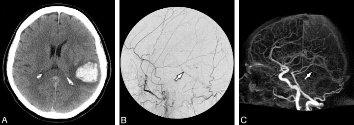
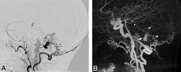
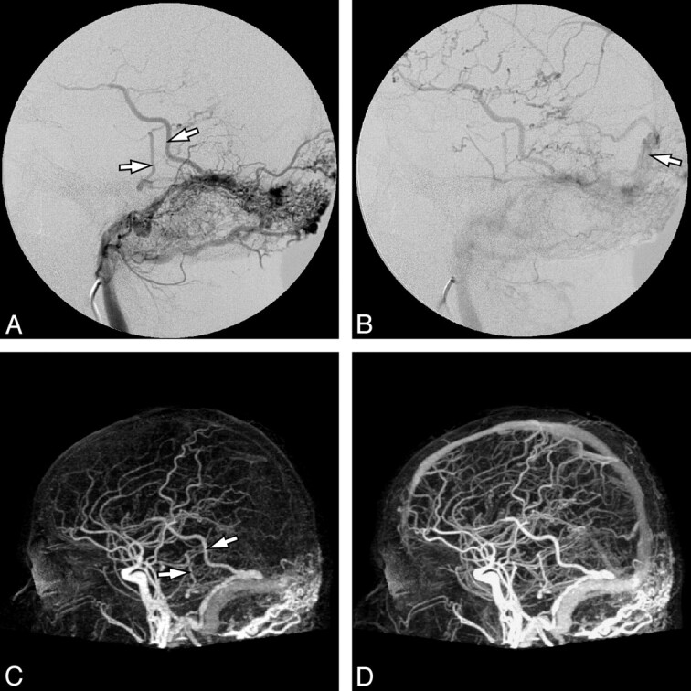
Background and purpose: The criterion standard to diagnose and classify cranial DAVFs is DSA. Since this is invasive, relatively expensive and time-consuming, a noninvasive alternative is of interest. We aimed to evaluate the capabilities and pitfalls of 4D-CTA in a consecutive series of patients who presented with a newly diagnosed cranial DAVF, as demonstrated by conventional DSA.
Materials and methods: Eleven patients were included in this study after biplane DSA demonstrated a cranial DAVF. They subsequently underwent 4D-CTA imaging by using a 320-detector CT scanner. DSA and 4D-CTA studies were independently read by 2 blinded observers, by using a standardized scoring sheet. 4D-CTA results were analyzed with DSA as the criterion standard.
Results: In 10 cases, there was full agreement between DSA and 4D-CTA regarding the Borden classification. However, in the remaining patient, a slow-filling DAVF with a low shunt volume was missed by both readers on 4D-CTA. In all 10 detected cases, ≥ 1 of the major contributing arteries could be identified with 4D-CTA. Although, by using DSA, the 2 observers identified additional arterial feeders in 7 and 8 cases, respectively, these discrepancies did not influence clinical decision making.
Conclusions: Although novel 4D-CTA imaging may not rule out a small slow-flow DAVF, it appears to be a valuable new adjunct in the noninvasive diagnostic work-up, treatment planning, and follow-up of patients with cranial DAVFs.
Figures
Similar articles
-
4D-CT angiography differentiating arteriovenous fistula subtypes.Clin Neurol Neurosurg. 2013 Aug;115(8):1313-6. doi: 10.1016/j.clineuro.2012.12.015. Epub 2013 Jan 5. Clin Neurol Neurosurg. 2013. PMID: 23298975
-
Four-dimensional computed tomography angiography is valuable in intracranial dural arteriovenous fistula diagnosis and fistula evaluation.Acta Neurol Belg. 2015 Sep;115(3):303-9. doi: 10.1007/s13760-014-0387-7. Epub 2014 Oct 30. Acta Neurol Belg. 2015. PMID: 25354667
-
High sensitivity and specificity of 4D-CTA in the detection of cranial arteriovenous shunts.Eur Radiol. 2019 Nov;29(11):5961-5970. doi: 10.1007/s00330-019-06234-4. Epub 2019 May 14. Eur Radiol. 2019. PMID: 31089848 Free PMC article.
-
Classification schemes of cranial dural arteriovenous fistulas.Neurosurg Clin N Am. 2012 Jan;23(1):55-62. doi: 10.1016/j.nec.2011.09.003. Neurosurg Clin N Am. 2012. PMID: 22107858 Review.
-
[Diagnostic imaging of cerebrovascular disease on multi-detector row computed tomography (MDCT)].Brain Nerve. 2011 Sep;63(9):923-32. Brain Nerve. 2011. PMID: 21878694 Review. Japanese.
Cited by
-
Whole-brain CT perfusion imaging using increased sampling intervals: A pilot study.Exp Ther Med. 2017 Sep;14(3):2643-2649. doi: 10.3892/etm.2017.4816. Epub 2017 Jul 19. Exp Ther Med. 2017. PMID: 28962207 Free PMC article.
-
Cerebral proliferative angiopathy depicted by four-dimensional computed tomographic angiography: A case report.Radiol Case Rep. 2022 May 5;17(7):2332-2336. doi: 10.1016/j.radcr.2022.03.104. eCollection 2022 Jul. Radiol Case Rep. 2022. PMID: 35570862 Free PMC article.
-
4D DSA for Dynamic Visualization of Cerebral Vasculature: A Single-Center Experience in 26 Cases.AJNR Am J Neuroradiol. 2017 Jun;38(6):1169-1176. doi: 10.3174/ajnr.A5161. Epub 2017 Apr 13. AJNR Am J Neuroradiol. 2017. PMID: 28408632 Free PMC article.
-
Optimization of the reconstruction interval in neurovascular 4D-CTA imaging. A technical note.Interv Neuroradiol. 2012 Dec;18(4):377-9. doi: 10.1177/159101991201800402. Epub 2012 Dec 3. Interv Neuroradiol. 2012. PMID: 23217631 Free PMC article.
-
Usefulness of 4D-CTA in the detection of cerebral dural sinus occlusion or stenosis with collateral pathways.Neuroradiol J. 2013 Aug;26(4):428-38. doi: 10.1177/197140091302600408. Epub 2013 Aug 27. Neuroradiol J. 2013. PMID: 24007731 Free PMC article.
References
-
- Cognard C, Gobin YP, Pierot L, et al. . Cerebral dural arteriovenous fistulas: clinical and angiographic correlation with a revised classification of venous drainage. Radiology 1995;194:671–80 - PubMed
-
- Lasjaunias P, Manelfe C, Chiu M.. Angiographic architecture of intracranial vascular malformations and fistulas: pretherapeutic aspects. Neurosurg Rev 1986;9:253–63 - PubMed
-
- Satomi J, van Dijk JM, terBrugge KG, et al. . Benign cranial dural arteriovenous fistulas: outcome of conservative management based on the natural history of the lesion. J Neurosurg 2002;97:767–70 - PubMed
-
- van Dijk JM, terBrugge KG, Willinsky RA, et al. . Clinical course of cranial dural arteriovenous fistulas with long-term persistent cortical venous reflux. Stroke 2002;33:1233–36 - PubMed
-
- Borden JA, Wu JK, Shucart WA.. A proposed classification for spinal and cranial dural arteriovenous fistulous malformations and implications for treatment. J Neurosurg 1995;82:166–79 - PubMed
Publication types
MeSH terms
LinkOut - more resources
Full Text Sources
Medical