

Treatment of brain arteriovenous malformations by double arterial catheterization with simultaneous injection of Onyx: retrospective series of 17 patients
- PMID: 20966066
- PMCID: PMC7964972
- DOI: 10.3174/ajnr.A2247
Treatment of brain arteriovenous malformations by double arterial catheterization with simultaneous injection of Onyx: retrospective series of 17 patients
Abstract
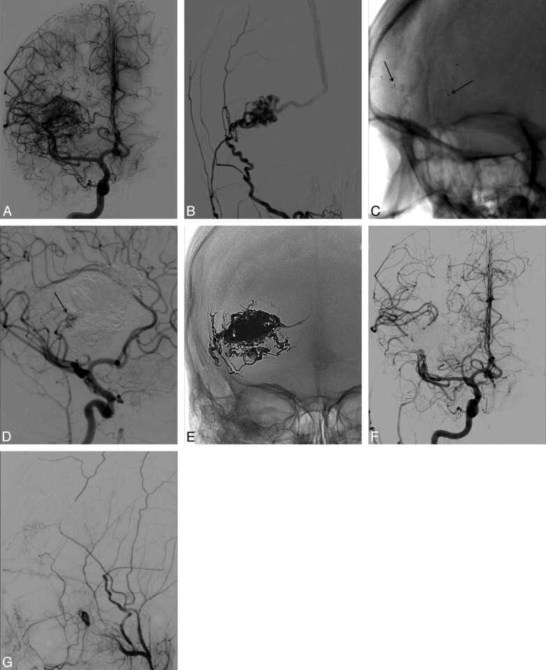
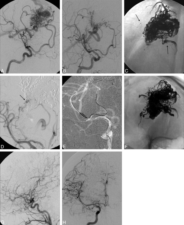
Background and purpose: The use of Onyx in the treatment of intracranial AVMs has increased the cure rate of endovascular embolization compared with the use of liquid adhesive agents. Inadvertent occlusion of the draining veins before the complete exclusion of the nidus constitutes a major risk of bleeding. We report a case series using the technique of double simultaneous arterial catheterization as an approach to achieve the complete exclusion of the nidus before reaching the venous drainage, through a more controlled hemodynamic filling.
Materials and methods: Between April 2008 and November 2009, 17 patients with brain AVMs were treated by the DACT. The mean age of the patients was 32.7 years (range, 6-54 years), with 9 females and 8 males. The clinical onset was characterized by intracranial hemorrhage in 8 patients and by seizures in 7. The size of the AVMs ranged from 13 to 54 mm (average, 26.2 mm). The DACT was always used with the objective of curing the AVM.
Results: All 17 patients completed the EVT. The average number of sessions conducted was 1.4 (range, 1-3 sessions), with the average injection amount of 6.9 mL of Onyx (range, 2-25.2 mL). Sixteen AVMs (94.1%) were angiographically cured by embolization. Clinical complications occurred in 2 patients (11.7%); 1 of these was permanent (5.9%). No deaths were registered.
Conclusions: This preliminary series shows that the DACT presents satisfactory results when used with curative intent.
Figures
Comment in
-
Comment on: treatment of brain arteriovenous malformations by double arterial catheterization with simultaneous injection of Onyx: retrospective series of 17 patients: Abud DG, Riva R, Nakiri GS, Padovani F, Khawaldeh M, Mounayer C. AJNR Am J Neuroradiol. 2011;32:152-158.Clin Neuroradiol. 2011 Jun;21(2):107-9. doi: 10.1007/s00062-011-0079-0. Clin Neuroradiol. 2011. PMID: 21656259 No abstract available.
Similar articles
-
Endovascular treatment of brain arteriovenous malformations with prolonged intranidal Onyx injection technique: long-term results in 350 consecutive patients with completed endovascular treatment course.J Neurosurg. 2011 Jul;115(1):78-88. doi: 10.3171/2011.2.JNS09830. Epub 2011 Apr 8. J Neurosurg. 2011. PMID: 21476804
-
Neurological morbidity and mortality associated with the endovascular treatment of cerebral arteriovenous malformations before and during the Onyx era.J Neurosurg. 2015 Jun;122(6):1492-7. doi: 10.3171/2015.2.JNS131368. Epub 2015 Mar 27. J Neurosurg. 2015. PMID: 25816081
-
Comment on: Curative Embolization of Brain Arteriovenous Malformations with Onyx: Patient Selection, Embolization Technique, and Results.Clin Neuroradiol. 2012 Jun;22(2):181-2. doi: 10.1007/s00062-012-0149-y. Epub 2012 May 8. Clin Neuroradiol. 2012. PMID: 22566010 Clinical Trial.
-
Transarterial coil-augmented Onyx embolization for brain arteriovenous malformation. Technique and experience in 22 consecutive patients.Interv Neuroradiol. 2014 Jan-Feb;20(1):83-90. doi: 10.15274/INR-2014-10012. Epub 2014 Feb 10. Interv Neuroradiol. 2014. PMID: 24556304 Free PMC article.
-
Long-term follow-up of transarterial balloon-assisted Onyx embolization for endovascular treatment of dural arteriovenous fistulas: A single-institution case series and literature review.Clin Neurol Neurosurg. 2020 Dec;199:106256. doi: 10.1016/j.clineuro.2020.106256. Epub 2020 Oct 1. Clin Neurol Neurosurg. 2020. PMID: 33069089 Review.
Cited by
-
Acute management of brain arteriovenous malformations.Curr Treat Options Neurol. 2015 May;17(5):346. doi: 10.1007/s11940-015-0346-5. Curr Treat Options Neurol. 2015. PMID: 25854649
-
ihtObtura: A novel liquid embolic agent with post-embolization radiopacity loss, in endovascular treatment of brain arteriovenous malformations, dural arteriovenous fistulas, and tumors: CLARIDAD trial.J Neurointerv Surg. 2024 Jul 16;17(5):493-499. doi: 10.1136/jnis-2023-021442. J Neurointerv Surg. 2024. PMID: 39019507 Free PMC article. Clinical Trial.
-
Transarterial Balloon-assisted Onyx Embolization of Intracranial Arteriovenous Malformations Using a Dual-lumen Balloon Microcatheter: Two Case Reports.J Cerebrovasc Endovasc Neurosurg. 2017 Sep;19(3):223-230. doi: 10.7461/jcen.2017.19.3.223. Epub 2017 Sep 30. J Cerebrovasc Endovasc Neurosurg. 2017. PMID: 29159158 Free PMC article.
-
Multiplug flow control technique as a novel transarterial curative approach for the endovascular treatment of cerebrovascular malformations.BMJ Case Rep. 2021 May 4;14(5):1-3. doi: 10.1136/bcr-2021-017418. BMJ Case Rep. 2021. PMID: 33947684 Free PMC article.
-
Primary endovascular embolisation of intracranial arteriovenous malformations (AVM)-UK single centre experience.Neuroradiology. 2024 Feb;66(2):227-236. doi: 10.1007/s00234-023-03258-y. Epub 2023 Nov 24. Neuroradiology. 2024. PMID: 37999787
References
-
- Richling B, Killer M, Al-Schameri AR, et al. . Therapy of brain arteriovenous malformations: multimodality treatment from a balanced standpoint. Neurosurgery 2006;59:S148–57 - PubMed
-
- Picard L, Moret J, Lepoire J, et al. . Endovascular treatment of intracerebral arteriovenous angiomas: technique, indications and results. J Neuroradiol 1984;11:9–28 - PubMed
-
- Katsaridis V, Papagiannaki C, Aimar E. Curative embolization of cerebral arteriovenous malformations (AVMs) with Onyx in 101 patients. Neuroradiology 2008;50:589–97 - PubMed
Publication types
MeSH terms
Substances
LinkOut - more resources
Full Text Sources
Miscellaneous