. 2010 Dec 24;285(52):41152-60.
doi: 10.1074/jbc.M110.158352. Epub 2010 Oct 21.

Phosphorylation of p50 NF-kappaB at a single serine residue by DNA-dependent protein kinase is critical for VCAM-1 expression upon TNF treatment

Affiliations

Phosphorylation of p50 NF-kappaB at a single serine residue by DNA-dependent protein kinase is critical for VCAM-1 expression upon TNF treatment

Jihang Ju et al. J Biol Chem. .

Abstract

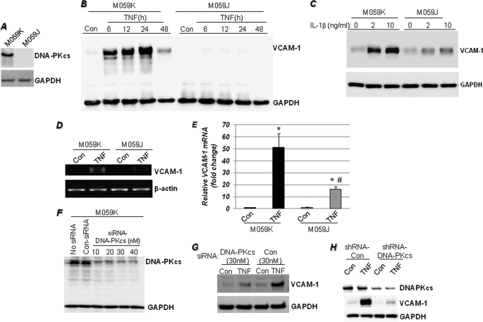
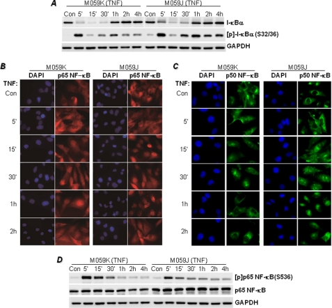
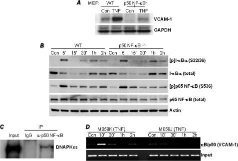
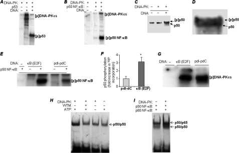
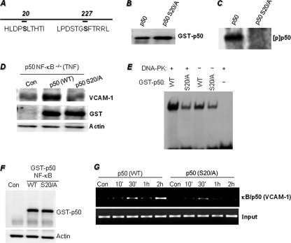
The DNA binding activity of NF-κB is critical for VCAM-1 expression during inflammation. DNA-dependent protein kinase (DNA-PK) is thought to be involved in NF-κB activation. Here we show that DNA-PK is required for VCAM-1 expression in response to TNF. The phosphorylation and subsequent degradation of I-κBα as well as the serine 536 phosphorylation and nuclear translocation of p65 NF-κB were insufficient for VCAM-1 expression in response to TNF. The requirement for p50 NF-κB in TNF-induced VCAM-1 expression may be associated with its interaction with and phosphorylation by DNA-PK, which appears to be dominant over the requirement for p65 NF-κB activation. p50 NF-κB binding to its consensus sequence increased its susceptibility to phosphorylation by DNA-PK. Additionally, DNA-PK activity appeared to increase the association between p50/p50 and p50/p65 NF-κB dimers upon binding to DNA and after binding of p50 NF-κB to the VCAM-1 promoter. Analyses of the p50 NF-κB protein sequence revealed that both serine 20 and serine 227 at the amino terminus of the protein are putative sites for phosphorylation by DNA-PK. Mutation of serine 20 completely eliminated phosphorylation of p50 NF-κB by DNA-PK, suggesting that serine 20 is the only site in p50 NF-κB for phosphorylation by DNA-PK. Re-establishing wild-type p50 NF-κB, but not its serine 20/alanine mutant, in p50 NF-κB(-/-) fibroblasts reversed VCAM-1 expression after TNF treatment, demonstrating the importance of the serine 20 phosphorylation site in the induction of VCAM-1 expression. Together, these results elucidate a novel mechanism for the involvement of DNA-PK in the positive regulation of p50 NF-κB to drive VCAM-1 expression.


Figures

FIGURE 1.
FIGURE 1.
DNA-PK is required for VCAM-1 expression in response to TNF treatment. A, immunoblot analysis of extracts from untreated M059K (DNA-PK-proficient) or M059J (DNA-PK-deficient) cells with antibodies to human DNA-PKcs or GAPDH. B, M059K and M059J cells were stimulated with TNF for different times, after which protein extracts were subjected to immunoblot analysis with antibodies to VCAM-1 or GAPDH. Con, control. C, M059K and M059J cells were stimulated with 2 or 10 ng/ml IL-1β for 18 h, after which protein extracts were subjected to immunoblot analysis with antibodies to VCAM-1 or GAPDH. D and E, cells were treated with TNF for 6 h, after which total RNA was prepared and subjected to cDNA generation followed by conventional (D) or quantitative (E) PCR with primers specific to human VCAM-1 or β-actin. *, different from respective untreated cells; #, different from TNF-treated M059K cells; p < 0.01. F, M059K cells were treated with different doses of DNA-PKcs siRNA; 48 h later, protein extracts were prepared and subjected to immunoblot analysis with antibodies to DNA-PKcs or GAPDH. G, M059K cells were treated with siRNA against DNA-PKcs or control siRNA; 48 h later, cells were treated with TNF for 18 h. Protein extracts were subjected to immunoblot analysis with antibodies to VCAM-1 or GAPDH. H, M059K cells were transduced with a lentiviral vector (Santa Cruz Biotechnology) expressing either control shRNA or an shRNA targeting DNA-PKcs. Forty-eight hours later, cells were treated with TNF for 18 h, and the resulting protein extracts were subjected to immunoblot analysis with antibodies to DNA-PKcs, VCAM-1, or GAPDH.
FIGURE 2.
FIGURE 2.
Effects of DNA-PK deficiency on I-κBα phosphorylation and its subsequent degradation and NF-κB phosphorylation and nuclear translocation in response to TNF stimulation. A, M059K and M059J cells were treated with TNF for the indicated times, after which protein extracts were subjected to immunoblot analysis with antibodies to I-κBα, phosphor(Ser-36)-I-κBα ([p]-IBα(S32/36)), or GAPDH. M059K and M059J cells grown in chamber slides were treated with TNF for the indicated times. Con, control. B and C, cells were then fixed and subjected to immunofluorescence staining with antibodies to p65 (B) or p50 NF-κB (C) followed by staining with DAPI (nuclei). D, the immunoblots displayed in A were stripped and reprobed with antibodies to the phosphorylated form of p 65 NF-κB at serine 536 ([p]p65NF-κB(S536)) or to antibodies recognizing full-length p65 NF-κB. Note that the immunoblot for GAPDH is the same as displayed in A.
FIGURE 3.
FIGURE 3.
Requirement of p50 NF-κB for TNF-induced VCAM-1 expression is dominant over the requirement for p65 NF-κB activation and may be associated with the interaction between p50 NF-κB and DNA-PK and the requirement for DNA-PK for efficient κB site occupancy on the VCAM-1 promoter by p50 NF-κB. A, MEFs derived from WT or p50−/− NF-κB mice were treated with 10 ng/ml TNF for 18 h. Protein extracts were subjected to immunoblot analysis with antibodies to VCAM-1 or GAPDH. Con, control. B, WT and p50−/− NF-κB MEFs were treated with TNF for the indicated times, after which protein extracts were subjected to immunoblot analysis with antibodies to I-κBα, phospho(Ser-32/36)-I-κBα ([p]-I-κBα(S32/36)), phospho(Ser-536)-p65 NF-κB ([p]p65 NF-κB (S536)), p65 NF-κB, or actin. C, M059K cells were treated with TNF for 1 h, after which nuclear extracts were prepared and subjected to immunoprecipitation (IP) with antibodies to DNA-PKcs or with normal IgG. Immunoprecipitates were subjected to immunoblot analysis with antibodies to DNA-PKcs. Ten percent of the amount used for immunoprecipitation was used as input control (see supplemental Fig. S2 for the original uncropped immunoblot). D, M059K or M059J cells were treated with TNF for the indicated times. Cells were then fixed and subjected to ChIP assay using antibodies to p50 NF-κB. Levels of immunoprecipitated chromatin fragments (−1015 to −775 region) of the human VCAM-1 promoter or input were examined by PCR.
FIGURE 4.
FIGURE 4.
Phosphorylation of p50 NF-κB by DNA-PK, effects of p50 NF-κB binding to a κB-containing DNA sequence on phosphorylation susceptibility, and consequences of p50 NF-κB phosphorylation on its DNA binding activity. A and B, in vitro kinase assay was performed using [32P]ATP and purified DNA-PK complex (Promega) with pure recombinant p53 (A), a positive control, or recombinant p50 NF-κB (B). The reactions were terminated by the addition of sample buffer and subjected to SDS-PAGE followed by autoradiography. [p] indicates phospho. C, the kinase reaction was repeated with cold ATP, and the reaction was subjected to immunoblot analysis with antibodies to p50 NF-κB; D represents a larger image of the double band for better visualization. E, recombinant p50 NF-κB was incubated in the presence of either a 500-bp κB-containing fragment of the E2F gene promoter or synthetic poly(dI-dC) for 15 min before the addition of DNA-PK and [32P]ATP in the kinase reactions. The reactions were terminated and subjected to SDS-PAGE followed by autoradiography. 32P incorporation in the p50 NF-κB bands was assessed using a Storm PhosphorImager system (GE Healthcare). pdI-pdC, poly(dI-dC). F, data are the means ± S.D. of values from three separate reactions. *, different from p50 NF-κB phosphorylated in a reaction containing poly(dI-dC); p < 0.01. G, autophosphorylation of DNA-PK in response to activation by either poly(dI-dC) or the 500-bp κB-containing fragment of the E2F gene promoter. H, p50 NF-κB was incubated in a kinase reaction in the presence or absence of DNA-PK, ATP (cold), and the DNA-PK inhibitor wortmannin (WTM) as shown in the figure. All kinase reactions were terminated by the addition of wortmannin. The reactions were then subjected to EMSA with a 32P end-labeled oligonucleotide containing the NF-κB-binding site. Gels were dried and subjected to autoradiography. I, p50 NF-κB was incubated in the presence or absence of DNA-PK in a kinase reaction. After reaction termination by wortmannin, p65 NF-κB was added prior to incubation with the 32P-labeled NF-κB oligonucleotide probe.
FIGURE 5.
FIGURE 5.
Phosphorylation of p50 NF-κB on a single serine residue (serine 20) at the amino terminus and requirement of this phosphorylation site for TNF-induced VCAM-1 expression. A, using NetPhosK 1.0 and NetPhos 2.0 software, two DNA-PK phosphorylation sites on p50 NF-κB at serines 20 and 227 were predicted. B, p50 NF-κB-tagged with GST was subjected to site-directed mutagenesis to achieve the substitution of alanine for serine 20. The generated expression vectors were transiently transfected into 293T cells. Purified WT or S20A mutant GST-tagged p50 NF-κB proteins were subjected to immunoblot analysis with antibodies to GST. C, purified WT or S20A mutant GST-tagged p50 NF-κB proteins were subjected to a kinase reaction with DNA-PK and [32P]ATP; the reactions were terminated by the addition of sample buffer and subjected to SDS-PAGE followed by autoradiography. [p] indicates phospho. D, p50−/− NF-κB MEFs were transfected with plasmids expressing WT or S20A mutant p50 (p50 S20/A) NF-κB or a control vector (Con) by electroporation. Cells were then treated with TNF for 18 h. The resulting protein extracts were subjected to immunoblot analysis with antibodies to VCAM-1, GST, or actin. E, purified WT or S20A mutant GST-tagged p50 NF-κB proteins were incubated in a kinase reaction in the presence or absence DNA-PK and ATP (cold). All kinase reactions were terminated by the addition of wortmannin. The reactions were then subjected to EMSA with a 32P end-labeled oligonucleotide containing the NF-κB-binding site. Gels were dried and subjected to autoradiography. F, p50−/− NF-κB MEFs were transfected with plasmids expressing WT or S20A mutant p50 NF-κB or a control vector (Con) by electroporation. Protein extracts from untreated cells were subjected to immunoblot with antibodies to GST or actin. G, a ChIP assay was performed using antibodies to p50 NF-κB in WT or S20A mutant p50-expressing p50−/− NF-κB MEFs treated with TNF for the indicated times. Levels of immunoprecipitated chromatin fragments (the −296 to −53 region) of the mouse VCAM-1 promoter or input were examined by PCR.

Similar articles

Cited by

References

    1. Braun M., Pietsch P., Schrör K., Baumann G., Felix S. B. (1999) Cardiovasc Res. 41, 395–401 - PubMed
    1. Hughes J. M., Arthur C. A., Baracho S., Carlin S. M., Hawker K. M., Johnson P. R., Armour C. L. (2000) Mediators Inflamm. 9, 93–99 - PMC - PubMed
    1. Makrilia N., Kollias A., Manolopoulos L., Syrigos K. (2009) Cancer Invest. 27, 1023–1037 - PubMed
    1. Rojas A. I., Ahmed A. R. (1999) Crit. Rev. Oral Biol. Med. 10, 337–358 - PubMed
    1. Huo Y., Ley K. (2001) Acta. Physiol. Scand. 173, 35–43 - PubMed

Publication types

MeSH terms