. 2010 Dec 31;285(53):41724-31.
doi: 10.1074/jbc.M110.156380. Epub 2010 Oct 21.

Functional dissection of adenylate cyclase R, an inducer of spore encapsulation

Affiliations

Functional dissection of adenylate cyclase R, an inducer of spore encapsulation

Zhi-hui Chen et al. J Biol Chem. .

Abstract

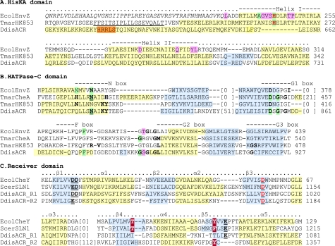
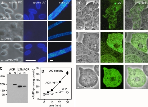
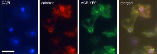
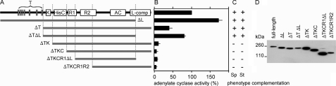
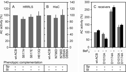
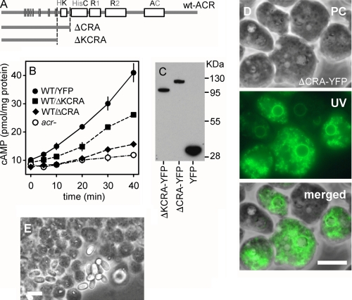
Cyclic AMP acting on protein kinase A controls sporulation and encystation in social and solitary amoebas. In Dictyostelium discoideum, adenylate cyclase R (ACR), is essential for spore encapsulation. In addition to its cyclase (AC) domain, ACR harbors seven transmembrane helices, a histidine kinase domain, and two receiver domains. We investigated the role of these domains in the regulation of AC activity. Expression of an ACR-YFP fusion protein in acr(-) cells rescued their sporulation defective phenotype and revealed that ACR is associated with the nuclear envelope and endoplasmic reticulum. Loss of the transmembrane helices (ΔTM) caused a 60% reduction of AC activity, but ΔTM-ACR still rescued the acr(-) phenotype. The isolated AC domain was properly expressed but inactive. Mutation of three essential ATP-binding residues in the histidine kinase domain did not affect the AC activity or phenotypic rescue. Mutation of the essential phosphoryl-accepting aspartate in receivers 1, 2, or both had only modest effects on AC activity and did not affect phenotypic rescue, indicating that AC activity is not critically regulated by phosphorelay. Remarkably, the dimerizing histidine phosphoacceptor subdomain, which in ACR lacks the canonical histidine for autophosphorylation, was essential for AC activity. Transformation of wild-type cells with an ACR allele (ΔCRA) that is truncated after this domain inhibited AC activity of endogenous ACR and replicated the acr(-) phenotype. Combined with the observation that the isolated AC domain was inactive, the dominant-negative effect of ΔCRA strongly suggests that the defunct phosphoacceptor domain acquired a novel role in enforcing dimerization of the AC domain.


Figures

FIGURE 1.
FIGURE 1.
Alignment of ACR functional domains with structurally resolved homologous domains. The location of α-helices (yellow) and β-sheets (blue highlight) were retrieved from the NCBI Molecular Modeling Database entries for all structurally resolved structures or predicted using PSIPRED (41) for the ACR domains. Green, essential for autokinase activity in CheA (29, 30) and for autokinase but not receiver-directed phosphatase activity in EnvZ; pink, essential for receiver-directed phosphatase but not autokinase activity in EnvZ (25, 31). A, HisKA domain. The ACR HisKA domain and flanking sequence from the seventh TM domain up to the HisC domain were aligned with the HisKA domains and corresponding regions of EcolEnvZ and TmarHK853 (42, 43). Red text, phosphoryl-accepting histidine. Orange, HRRLS motif. Italic letters, HAMP domain; underlined letters, TM domain. Protein Data Bank codes are as follows: 1JOY, EcolEnvZ; 2C2A, TmarHK953. B, HATPase-C domain. The ACR HisC domain was aligned with three structurally resolved bacterial HisC domains. Residues in boldface type represent those demonstrated to interact with ATP (43–46); bold/underlined residues are those that interact with Mg2+ (45). When functionally essential residues are conserved in ACR, they are similarly marked. Protein Data Bank codes are as follows: 1BXD, EcolEnvZ; 1I59, TMarCheA; 2C2A, TmarHK853. C, receiver domain. The two ACR receiver domains R1 and R2 were aligned with two structurally resolved R domains from yeast and Escherichia coli. Red text, phosphoryl-accepting aspartate. Bold/underlined text, residues in the Mg2+-binding active site, maroon, switch residues that reorient upon phosphorylation (47–50). Protein Data Bank codes are as follows: 1F4V, EcolCheY; 2R25, ScerSLN1.
FIGURE 2.
FIGURE 2.
Expression and activity of a full-length ACR construct. A, complementation of acr. Full-length ACR cDNA was cloned into vector pB17S-EYFP in between the constitutive A15 promoter and YFP. This vector was transformed into an acr mutant to yield acr/ACR-YFP cells. Wild-type, acr/ACR-YFP, and acr cells transformed with empty vector (acr/YFP) were starved on PB agar until fruiting bodies had formed, which were transferred to a droplet of PB with 0.01% Calcofluor on a glass slide and photographed using a Leica DMLB2 fluorescence microscope under phase contrast (PC) and UV illumination. B, cellular localization of ACR. Phase contrast images and optical sections of YFP fluorescence in living acr/ACR-YFP, acr/YFP and acrTMACR-YFP cells were obtained with a Leica DMRBE confocal laser scanning microscope. C, cell fractionation. Vegetative acr/ACR-YFP and acrTMACR-YFP cells were lysed through nuclepore filters. Lysates were fractionated by differential centrifugation into cytoplasm (C) and nuclei (N) as described previously (51), and both fractions were subjected to qualitative Western blotting with α-GFP antibodies. D, cAMP production by ACR. Vegetative acr/ACR-YFP and acr/YFP cells were filter-lysed and incubated for 30 min at 22 °C with AC assay mix (see “Experimental Procedures”). cAMP accumulation was assayed at the indicated time periods and standardized on the protein content of the cell lysates. Means and S.D. of four experiments performed in triplicate are presented. Scale bars in A and B, 10 μm.
FIGURE 3.
FIGURE 3.
Visualization of ACR-YFP and calnexin by immunofluorescence. Axenically grown acr/ACR-YFP cells were harvested in exponential phase and triple stained with (i) a polyclonal rabbit-anti-GFP antibody, followed by FITC conjugated donkey anti-rabbit IgG; (ii) a monoclonal mouse-anti-calnexin antibody (24) followed by Alexa Fluor® 594 conjugated goat anti-mouse IgG; and (iii) DAPI to detect ACR-YFP, calnexin, and DNA, respectively. Cells were photographed through the UV, TRITC, and FITC filter sets of a Leica DMLB2 fluorescence microscope. The merged image was prepared with the Qcapture Pro camera software. Scale bar, 10 μm.
FIGURE 4.
FIGURE 4.
Functions of the different domains of ACR in adenylate cyclase regulation. A, schematic of truncations. Truncated segments of the ACR cDNA lacking transmembrane (T, vertical bars), HisKA (K), HisC (C), receiver (R1 and R2) domains or the C-terminal low complexity region (L) were prepared by PCR amplification and recombined with unaltered segments in pGEM7ZF+ to reconstitute the entire (but truncated) cDNAs, which were then transferred to vector pB17S-EYFP and transformed into acr cells. B, AC activity of truncated proteins. Lysates of transformed cell lines were incubated with AC assay mix for 30 min and assayed for cAMP production. Data are standardized on the amount of YFP fusion protein in the lysates as determined by quantitative Western blotting (see “Experimental Procedures”). The results represent means and S.D. of four experiments performed on cells from at least two separate transformations. C, complementation of acr phenotype. All transformants were developed into fruiting bodies, which were examined as described for Fig. 2A for complementation of the spore encapsulation (Sp) and thin stalk (St) phenotype. D, expression levels of truncated proteins. Lysates of 3 × 10⁵ cells that had been transformed with intact and truncated ACR-YFP fusion constructs were size-fractionated by SDS-PAGE. The YFP-fusion proteins were visualized by qualitative Western blotting using an α-GFP antibody.
FIGURE 5.
FIGURE 5.
Disruption of domain activity by site-directed mutagenesis. A, putative phosphoryl-accepting residues in the HRRLS motif. His613 in the HRRLS motif, which resides in segment AB of ACR (supplemental Fig. S1B), was mutated into Gln, whereas Ser617 was mutated either into Ala or Glu. The mutated segments were recombined with segments CD, EF, and GH to recreate full-length ACR-YFP, which was transformed into acr cells and assayed for AC activity as described above. Data are expressed as percentage of AC activity in intact ACR. The transformed acr cells were additionally developed into fruiting bodies to examine complementation of their spore- and stalk maturation phenotypes by the constructs. B, residues essential for histidine kinase activity. Asn769 in the HisC domain was first mutated into Asp in segment CD. Later, Glu854 and Gly856 were both mutated into Ala in segment CD that contained the N769D mutation. The two mutated segments were recombined with segments AB, EF, and GH to recreate full-length ACR-YFP proteins, transformed into acr cells, and assayed for AC activity and complementation of the acr phenotype. C, receiver domains. The phosphoryl-accepting residues Asp1010 in R1 and Asp1174 in R2 were mutated into Ala in the individual ACR segments CD and EF, respectively, or in both. The mutated segments were recombined with complementary segments to recreate full-length ACR-YFP and transformed into acr cells. AC activity was assayed in the presence 5 mm NaF (−) or 5 mm NaF with 0.1 mm BeCl2 to form BeF3 (+) (34). Data represent means and S.E. of three experiments performed in triplicate. Sp, spore encapsulation; St, thin stalk.
FIGURE 6.
FIGURE 6.
Effects of N-terminal ACR fragments on wild-type ACR activity. A, schematic of ACR constructs that are C-terminally truncated after the HisKA (ΔCRA) or transmembrane domains (ΔKCRA). The fragments were fused to YFP in pB17S-EYFP and transformed into wild-type cells. B, lysates of vegetative acr cells and wild-type cells transformed with ΔCRA-YFP, ΔKCRA-YFP, or empty vector (YFP) were assayed for cAMP synthesis under ACR assay conditions, which was standardized on the total protein content of the lysates. Means and S.E. of three experiments performed in triplicate are shown. C, aliquots of 15 μg of total protein of the transformed wild-type cell lines were subjected to qualitative Western blotting with α-GFP antibodies. D, visualization of ΔCRA-YFP localization in living vegetative cells by fluorescence and phase contrast microscopy. E, squashed spore heads of 2-day-old fruiting bodies of wild-type cells transformed with the ΔCRA-YFP construct contain mainly amoeboid cells. Scale bar, 10 μm. PC, phase contrast.

Similar articles

Cited by

References

    1. Linder J. U., Schultz J. E. (2003) Cellular Signalling 15, 1081–1089 - PubMed
    1. Ritchie A. V., van Es S., Fouquet C., Schaap P. (2008) Mol. Biol. Evol. 25, 2109–2118 - PMC - PubMed
    1. Baker D. A., Kelly J. M. (2004) Trends Parasitol. 20, 227–232 - PubMed
    1. Chen Y., Cann M. J., Litvin T. N., Iourgenko V., Sinclair M. L., Levin L. R., Buck J. (2000) Science 289, 625–628 - PubMed
    1. Kim H. J., Chang W. T., Meima M., Gross J. D., Schaap P. (1998) J. Biol. Chem. 273, 30859–30862 - PubMed

Publication types

LinkOut - more resources