. 2010 Oct 22;330(6003):509-12.
doi: 10.1126/science.1191750.

Insight into the mechanism of the influenza A proton channel from a structure in a lipid bilayer

Affiliations

Insight into the mechanism of the influenza A proton channel from a structure in a lipid bilayer

Mukesh Sharma et al. Science. .

Abstract

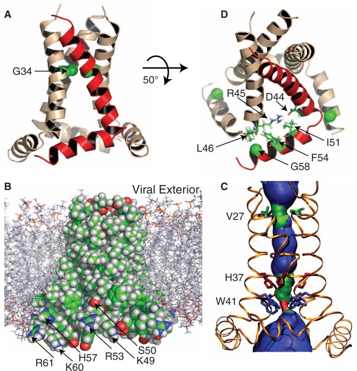
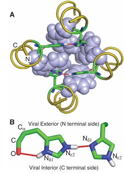
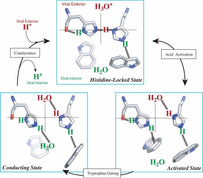
The M2 protein from the influenza A virus, an acid-activated proton-selective channel, has been the subject of numerous conductance, structural, and computational studies. However, little is known at the atomic level about the heart of the functional mechanism for this tetrameric protein, a His(37)-Trp(41) cluster. We report the structure of the M2 conductance domain (residues 22 to 62) in a lipid bilayer, which displays the defining features of the native protein that have not been attainable from structures solubilized by detergents. We propose that the tetrameric His(37)-Trp(41) cluster guides protons through the channel by forming and breaking hydrogen bonds between adjacent pairs of histidines and through specific interactions of the histidines with the tryptophan gate. This mechanism explains the main observations on M2 proton conductance.


Figures

Fig. 1
Fig. 1
The tetrameric structure of the M2 conductance domain, solved by solid-state NMR spectroscopy and restrained molecular dynamics simulations, in liquid crystalline lipid bilayers. See (32) for details and fig. S3 for the NMR spectra. (A) Ribbon representation of the TM and amphipathic helices. One monomer is shown in red. The TM helix is kinked around the highly conserved Gly34 (shown as Cα spheres). (B) Space-filling representation of the protein side chains in the lipid bilayer environment used for the NMR spectroscopy, structural refinement, and functional assay. C, O, N, and H atoms are colored green, red, blue, and white, respectively. The nonpolar residues of the TM and amphipathic helices form a continuous surface; the positively charged residues of the amphipathic helix are arrayed on the outer edge of the structure in optimal position to interact with charged lipids. The Ser50 hydroxyl is also shown to be in an optimal position (as Cys50) to accept a palmitoyl group in native membranes. (C) HOLE image (33) illustrating pore constriction at Val27 and Trp41. (D) Several key residues at the junction between the TM and amphipathic helices, including Gly58 (shown as Cα spheres), which facilitates the close approach of adjacent monomers, and Ile51 and Phe54, which fill a pocket previously described as a rimantadine-binding site (12).
Fig. 2
Fig. 2
The structure of the HxxxW quartet in the histidine-locked state. (A) Top view of the tetrameric cluster of H37xxxW41 (His37 as sticks and Trp41 as spheres). Note the near-coplanar arrangement of each imidazole-imidazolium dimer that forms a strong hydrogen bond between Nδ1 and Nε2. In each dimer, the remaining Nε2 interacts with the indole of a Trp41 residue through a cation-π interaction. The backbones have four-fold symmetry, as defined by the time-averaged NMR data. (B) Side view of one of the two imidazole-imidazolium dimers. Both the intraresidue Nδ1-H-O hydrogen bond and the interresidue Nε2-H-Nδ1 strong hydrogen bond can be seen. The near-linearity of the interresidue hydrogen bond is obtained at the expense of a strained Cα-Cβ-Cγ angle (enlarged by ~10°) of the residue on the left.
Fig. 3
Fig. 3
Proposed mechanism of acid activation and proton conductance illustrated with half of the HxxxW quartet from a side view. The histidine-locked state (top) is shown with a hydronium ion waiting in the N-terminal pore. Acid activation is initiated with a proton transfer from the hydronium ion into the interresidue hydrogen bond between Nδ1 and Nε2. In the resulting activated state, the two imidazolium rings rotate so that the two nitrogens move toward the center of the pore; in addition, the protonated Nδ1 forms a hydrogen bond with water in the N-terminal pore while the protonated Nε2 moves downward (via relaxing the Cα-Cβ-Cγ angle) to form a cation-π interaction with an indole, thereby blocking water access from the C-terminal pore. The conducting state is obtained when this indole moves aside to expose the Nε2 proton to a water in the C-terminal pore. It was suggested previously (27) that the indole motion involves ring rotation coupled to backbone kinking. Once the Nε2 proton is released to C-terminal water, the HxxxW quartet returns to the histidine-locked state.

Comment in

References

    1. Takeda M, Pekosz A, Shuck K, Pinto LH, Lamb RA. J. Virol. 2002;76:1391. - PMC - PubMed
    1. Gubareva L, et al. Centers for Disease Control and Prevention (CDC) Morb. Mortal. Wkly. Rep. 2009;58:1. - PubMed
    1. Nishimura K, Kim S, Zhang L, Cross TA. Biochemistry. 2002;41:13170. - PubMed
    1. Hu J, et al. Biophys. J. 2007;92:4335. - PMC - PubMed
    1. Stouffer AL, et al. Nature. 2008;451:596. - PMC - PubMed

Publication types

Associated data