. 2010 Nov;2(11):962-6.
doi: 10.1038/nchem.858. Epub 2010 Oct 3.

Recognition-mediated activation of therapeutic gold nanoparticles inside living cells

Affiliations

Recognition-mediated activation of therapeutic gold nanoparticles inside living cells

Chaekyu Kim et al. Nat Chem. 2010 Nov.

Abstract

Supramolecular chemistry provides a versatile tool for the organization of molecular systems into functional structures and the actuation of these assemblies for applications through the reversible association between complementary components. Use of this methodology in living systems, however, represents a significant challenge owing to the chemical complexity of cellular environments and lack of selectivity of conventional supramolecular interactions. Herein, we present a host-guest system featuring diaminohexane-terminated gold nanoparticles (AuNP-NH(2)) and complementary cucurbit[7]uril (CB[7]). In this system, threading of CB[7] on the particle surface reduces the cytotoxicity of AuNP-NH(2) through sequestration of the particle in endosomes. Intracellular triggering of the therapeutic effect of AuNP-NH(2) was then achieved through the administration of 1-adamantylamine (ADA), removing CB[7] from the nanoparticle surface, causing the endosomal release and concomitant in situ cytotoxicity of AuNP-NH(2). This supramolecular strategy for intracellular activation provides a new tool for potential therapeutic applications.


Figures
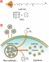
Figure 1
Figure 1. Structure of gold nanoparticle and use of intracellular host-guest complexation to trigger nanoparticle cytotoxicity
(a) Structure of diaminohexane-terminated gold nanoparticle (AuNP-NH2) and cucurbit[7]uril (CB[7]). (b) Activation of AuNP-NH2-CB[7] cytotoxicity by dethreading of CB[7] from the nanoparticle surface by ADA.
Figure 2
Figure 2. NMR titration and TEM of AuNP-NH2-CB[7] complex
(a) The resonance signals for the methylene groups (• and ★) of AuNP-NH2-CB[7] were shifted upfield, relatively to those in AuNP-NH2. (b) TEM image of AuNP-NH2-CB[7] complex. TEM sample was prepared by placing the desired AuNP-NH2-CB[7] solution (3 μM) on to a Cu grid coated with carbon film, followed by 2 % of uranyl acetate staining for 15 min. Increase of electron density of organic layer on nanoparticle after binding with CB[7] afford the organic shell of AuNP-NH2-CB[7] enough to be visualized in the TEM image. Organic shell on AuNP-NH2 was not observed in the same TEM sample preparation.
Figure 3
Figure 3. Cellular uptake of the gold nanoparticles
Quantification of the amount of gold present in cells. Samples were analyzed by ICP-MS to determine the amount of gold in MCF-7 cell after 3 h incubation with 0.5 μM of AuNP-NH2 and AuNP-NH2-CB[7]. Both particles showed almost same cellular uptake. Cellular uptake experiments with each gold nanoparticle were repeated 3 times, and each replicate was measured 5 times by ICP-MS. Error bars represent the standard deviations of these measurements.
Figure 4
Figure 4. Intracellular localization of the gold nanoparticles
TEM images of cross sectional MCF-7 cells incubated for 24 h with 2 μM (a) AuNP-NH2 and (b) AuNP-NH2-CB[7]. Significant amount of AuNP-NH2 is present in the cytosol, however most of the AuNP-NH2-CB[7] seems to be trapped in organelles such as endosome. (c) TEM images of cross sectional MCF-7 cells incubated for 3 h with 2 μM of AuNP-NH2-CB[7] and then further incubation with ADA for 24 h. In the intracellular environment ADA transforms AuNP-NH2-CB[7] to AuNP-NH2, which then escaped from the endosome and observed to be dispersed in the cytosol. i, ii and iii are the magnified sections from the first panel of part (c).
Figure 5
Figure 5. Cytotoxicity of AuNP-NH2 and AuNP-NH2-CB[7] and modulating cytotoxicity of the gold nanoparticles
(a) Cytotoxicity of AuNP-NH2 and AuNP-NH2-CB[7] measured by Alamar blue assay after 24 h incubation in MCF-7. IC50 of AuNP-NH2 was 1.3 μM and no cytotoxicity of AuNP-NH2-CB[7] was observed up to 50 μM. (b) Triggering cytotoxicity using ADA. After 3h incubation of AuNP-NH2-CB[7] (2 μM) in MCF-7 cell, different concentrations (0, 0.2 and 0.4 mM) of ADA in medium added and further incubated at 37 °C for 24 h. The cell viability was then determined by using an Alamar blue assay. As controls, cell viability of AuNP-NH2 and AuNP-NH2-CB[7] was measured after 24 h incubation (34 % and 100%, respectively). Cell viability experiments were performed as triplicate and the error bars represent the standard deviations of these measurements.

References

    1. Lehn JM. Toward self-organization and complex matter. Science. 2002;295:2400–2403. - PubMed
    1. Lehn JM. From supramolecular chemistry towards constitutional dynamic chemistry and adaptive chemistry. Chem. Soc. Rev. 2007;36:151–160. - PubMed
    1. Reinhoudt DN, Crego-Calama M. Synthesis beyond the molecule. Science. 2002;295:2403–2407. - PubMed
    1. Yaghi OM, et al. Reticular synthesis and the design of new materials. Nature. 2003;423:705–714. - PubMed
    1. Lehn JM. Supramolecular Chemistry: Concepts and Perspectives. VCH; New York: 1995.

Publication types