

Cystic macular oedema on spectral-domain optical coherence tomography in choroideremia patients without cystic changes on fundus examination
- PMID: 20966974
- PMCID: PMC3020991
- DOI: 10.1038/eye.2010.157
Cystic macular oedema on spectral-domain optical coherence tomography in choroideremia patients without cystic changes on fundus examination
Abstract
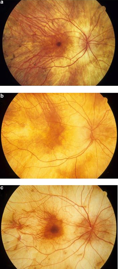
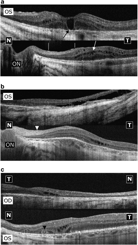
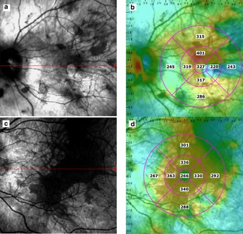
Purpose: To determine the prevalence of cystic macular oedema (CME) in patients with choroideremia (CHM) by using spectral-domain optical coherence tomography (SD-OCT).
Methods: A total 16 patients affected with CHM were enrolled in the study. All patients underwent a complete eye examination. SD-OCT was performed using an OPKO spectral-domain OCT/SLO instrument.
Results: The average age of the study patients was 44.0 ± 16.0 years (range, 13-63 years). Out of the 16 patients with CHM, 10 patients (62.5%) showed a degree of CME on SD-OCT testing in at least one eye, and 8 patients (50%) showed CME in both eyes.
Conclusions: Because of its notable prevalence, it would seem prudent to screen CHM patients by SD-OCT for the possible presence of CME and to identify those amenable to future treatment strategies for their macular oedema.
Figures
References
-
- McCulloch C, McCulloch RJP. A hereditary and clinical study of choroideremia. Trans Am Acad Ophthalmol Otolaryngol. 1948;52:160–190. - PubMed
-
- Renner AB, Kellner U, Cropp E, Preising MN, MacDonald IM, van den Hurk JA, et al. Choroideremia: variability of clinical and electrophysiological characteristics and first report of a negative electroretinogram. Ophthalmology. 2006;113:2066–2073. - PubMed
-
- Potter MJ, Wong E, Szabo SM, McTaggart KE. Clinical findings in a carrier of a new mutation in the choroideremia gene. Ophthalmology. 2004;111:1905–1909. - PubMed
-
- Majid MA, Horsborough B, Gray RH. Unusal macular findings in known choroideremia carrier. Eye (Lond) 1998;12:740–741. - PubMed
Publication types
MeSH terms
Grants and funding
LinkOut - more resources
Full Text Sources
