

doi: 10.1364/OL.35.003315.
Intraoperative spectral domain optical coherence tomography for vitreoretinal surgery
Affiliations
- PMID: 20967051
- PMCID: PMC3123722
- DOI: 10.1364/OL.35.003315
Item in Clipboard
Intraoperative spectral domain optical coherence tomography for vitreoretinal surgery
Opt Lett.
.
Abstract
We demonstrate in vivo human retinal imaging using an intraoperative microscope-mounted optical coherence tomography system (MMOCT). Our optomechanical design adapts an Oculus Binocular Indirect Ophthalmo Microscope (BIOM3), suspended from a Leica ophthalmic surgical microscope, with spectral domain optical coherence tomography (SD-OCT) scanning and relay optics. The MMOCT enables wide-field noncontact real-time cross-sectional imaging of retinal structure, allowing for SD-OCT augmented intrasurgical microscopy for intraocular visualization. We experimentally quantify the axial and lateral resolution of the MMOCT and demonstrate fundus imaging at a 20Hz frame rate.
Figures
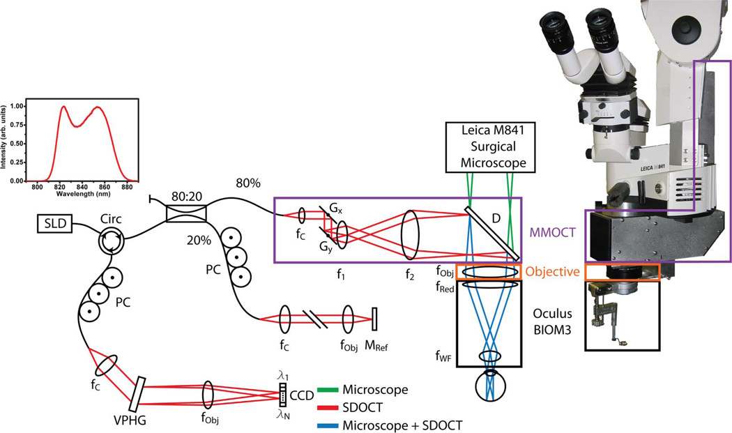
(Color online) Optical schematic and photograph of the MMOCT showing surgical microscope (green), SD-OCT (red), and shared (blue) optical paths. The sample-arm optics of the MMOCT (purple box) consists of galvanometer scanners, a relay telescope, a dichroic beam splitter, and focusing optics from the surgical microscope, including a microscope objective (orange box) and reduction and wide-field ophthalmic lenses (black box). CCD, linear CCD array; D, dichroic mirror; f, focal length of collimating, relay, and focusing elements; G, galvanometer; M, mirror; PC, polarization controller; VPHG, grating.
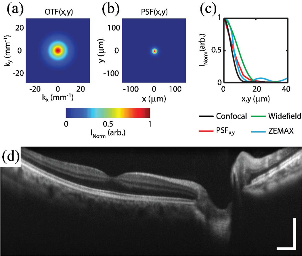
(Color online) Resolution of MMOCT. Focal plane cross sections of the lateral (a) OTF and (b) PSF, calculated from the Fourier transform of the OTF. (c) Lateral PSF cross section compared with theoretical values for confocal and wide-field imaging systems, and ZEMAX simulations. (d) Ten coregistered and averaged image excerpts from a video of 8 mm × 1.75 mm (lateral × depth) B-scans of in vivo human macula (Media 1 ). Images were acquired with 1024 × 1024 pixels (lateral × spectral) at a frame rate of 20 Hz. Illumination power, 700 µW; scale bar, 2°.
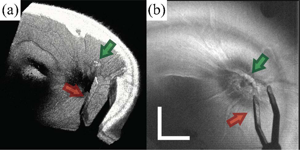
(Color online) MMOCT of surgical instrument (forceps) over the optic nerve in cadaveric porcine eye. The surgical procedure was performed by viewing through the surgical microscope with concurrent acquisition of MMOCT volumes. A 6 mm × 6 mm volumetric dataset was acquired with 500 B-scans, sampled with 1024 × 500 pixels (spectral × lateral) at a 20 kHz line rate. (a) Volumetric rendering (Media 2 ) and (b) SVP show both the instrument (red arrow) and a piece of glial tissue extruding from the optic nerve (green arrow). Illumination power, 700 µW; scale bar, 3°.
References
-
- Virata SR, Kylstra JA, Singh HT. Retina. 1999;19:287. - PubMed
-
- Charles S. Retina. 2008;28:1. - PubMed
-
- Ando F, Sasano K, Ohba N, Hirose H, Yasui O. Am. J. Ophthalmol. 2004;137:609. - PubMed
-
- Nassif NA, Cense B, Park BH, Pierce MC, Yun SH, Bouma BE, Tearney GJ, Chen TC, de Boer JF. Opt. Express. 2004;12:367. - PubMed
-
- Stopa M, Bower BA, Davies E, Izatt JA, Toth CA. Retina. 2008;28:298. - PubMed
Publication types
MeSH terms
Grants and funding
LinkOut - more resources
Full Text Sources
Other Literature Sources
Medical
