

doi: 10.1364/OL.35.003345.
Tomographic molecular imaging of x-ray-excitable nanoparticles
Affiliations
- PMID: 20967061
- PMCID: PMC12121645
- DOI: 10.1364/OL.35.003345
Item in Clipboard
Tomographic molecular imaging of x-ray-excitable nanoparticles
Opt Lett.
.
Abstract
X-ray luminescence computed tomography (XLCT) is proposed as a new dual molecular/anatomical imaging modality. XLCT is based on the selective excitation and optical detection of x-ray-excitable nanoparticles. As a proof of concept, we built a prototype XLCT system and imaged near-IR-emitting Gd(2)O(2)S:Eu phosphors in various phantoms. Imaging in an optically diffusive medium shows that imaging performance is not affected by optical scatter; furthermore, the linear response of the reconstructed images suggests that XLCT is capable of quantitative imaging.
Figures
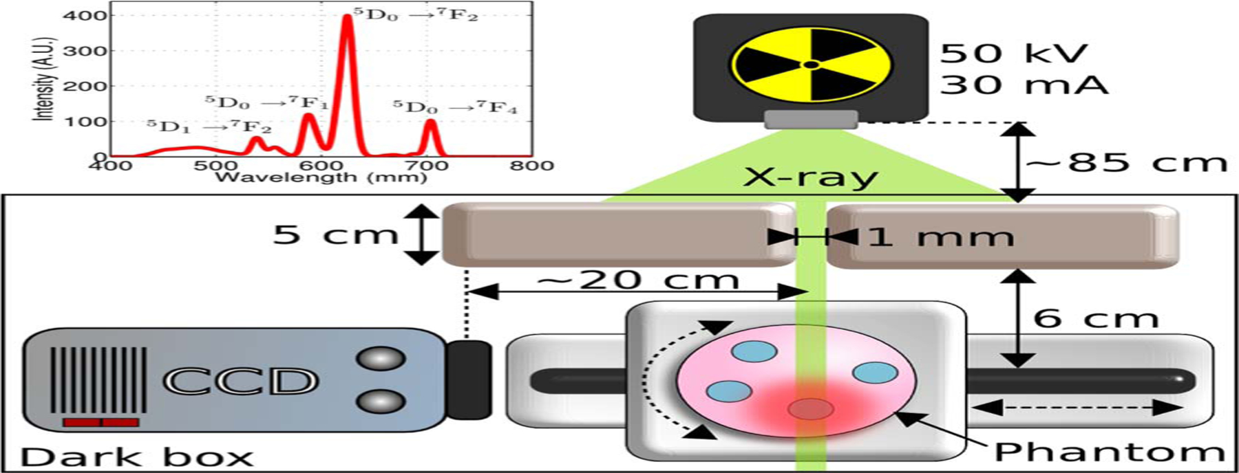
(Color online) A phantom containing phosphor inclusions is moved on a rotation/translation stage while being irradiated by a narrow, stationary x-ray beam. At each position, the XL signal was measured with an EM-CCD camera. Inset, XL spectrum for GOSE.
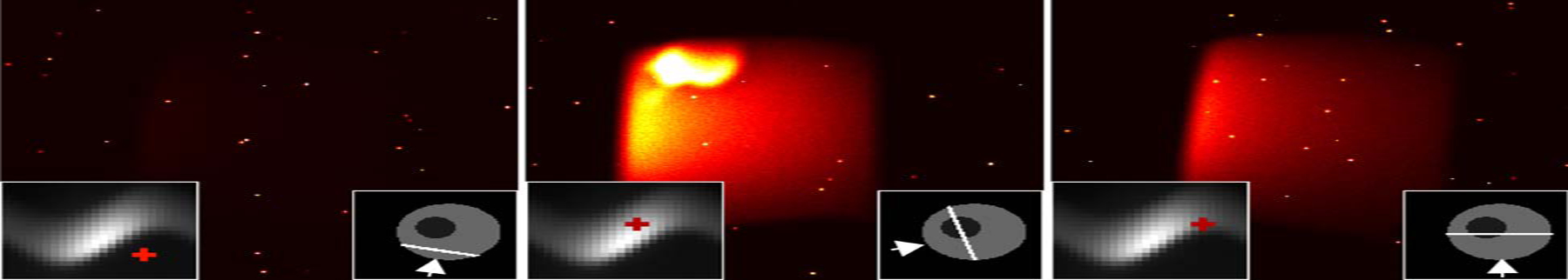
(Color online) Sample images acquired with an EMCCD camera (white arrow) of the turbid phantom under three different x-ray irradiations (bottom-right inset). Each image maps to a sinogram bin (red cross, bottom-left inset). The hot spot in the middle image was caused by a defect in the phantom.
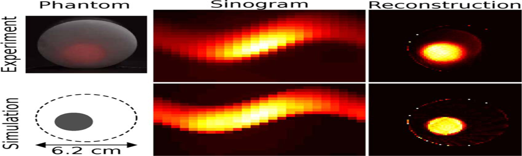
(Color online) A turbid phantom (left), composed of phosphor suspended in an optically diffusive cylinder, was acquired using XLCT to produce a sinogram (middle), which was reconstructed with 100 iterations of ML-EM (right). The simulation (bottom row) modeled the light propagation in a diffuse medium.

(Color online) A gradient phantom (left), composed of various phosphor concentrations embedded in a gel, was acquired using XLCT to produce a sinogram (middle), which was reconstructed with 100 iterations of ML-EM (right).

Response linearity for all seven phosphor inclusions, shown for the experimental and simulated gradient phantom.
References
-
- Speck U, in Molecular Imaging I (Springer, 2008), pp. 167–175.
-
- Xing M, Cao W, Pang T, Ling X, and Chen N, Chin. Sci. Bull 54, 2982 (2009).
-
- Tian Y, Cao W-H, Luo X-X, and Fu Y, J. Alloys Compd 433, 313 (2007).
-
- Sun C, Carpenter C, Pratx G, and Xing L, in World Molecular Imaging Congress (2010).
Publication types
MeSH terms
Substances
Grants and funding
LinkOut - more resources
Full Text Sources
Other Literature Sources
Medical
Research Materials
