. 2010 Oct 13;5(10):e13179.
doi: 10.1371/journal.pone.0013179.

Rapid and highly sensitive detection of malaria-infected erythrocytes using a cell microarray chip

Affiliations

Rapid and highly sensitive detection of malaria-infected erythrocytes using a cell microarray chip

Shouki Yatsushiro et al. PLoS One. .

Abstract

Background: Malaria is one of the major human infectious diseases in many endemic countries. For prevention of the spread of malaria, it is necessary to develop an early, sensitive, accurate and conventional diagnosis system.

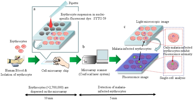
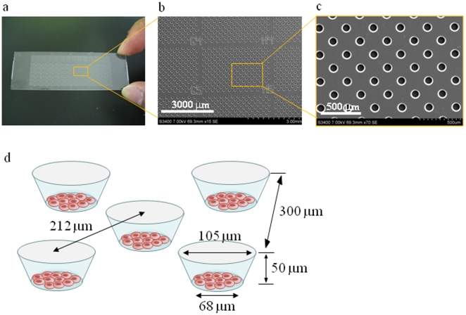
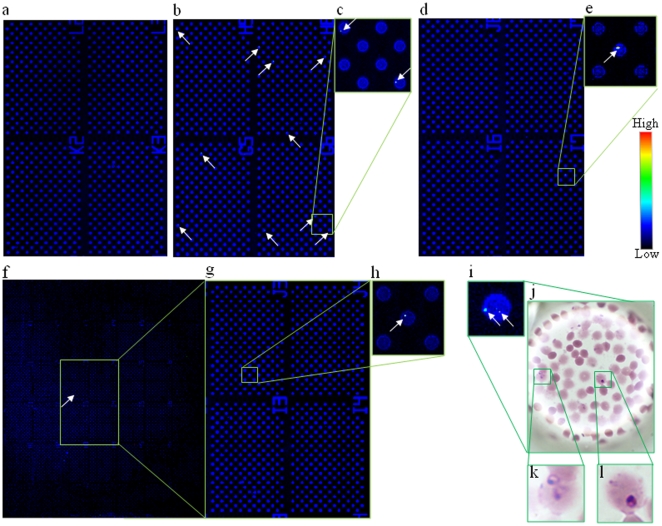
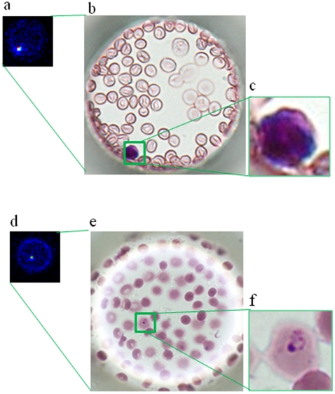
Methods and findings: A cell microarray chip was used to detect for malaria-infected erythrocytes. The chip, with 20,944 microchambers (105 µm width and 50 µm depth), was made from polystyrene, and the formation of monolayers of erythrocytes in the microchambers was observed. Cultured Plasmodium falciparum strain 3D7 was used to examine the potential of the cell microarray chip for malaria diagnosis. An erythrocyte suspension in a nuclear staining dye, SYTO 59, was dispersed on the chip surface, followed by 10 min standing to allow the erythrocytes to settle down into the microchambers. About 130 erythrocytes were accommodated in each microchamber, there being over 2,700,000 erythrocytes in total on a chip. A microarray scanner was employed to detect any fluorescence-positive erythrocytes within 5 min, and 0.0001% parasitemia could be detected. To examine the contamination by leukocytes of purified erythrocytes from human blood, 20 µl of whole blood was mixed with 10 ml of RPMI 1640, and the mixture was passed through a leukocyte isolation filter. The eluted portion was centrifuged at 1,000×g for 2 min, and the pellet was dispersed in 1.0 ml of medium. SYTO 59 was added to the erythrocyte suspension, followed by analysis on a cell microarray chip. Similar accommodation of cells in the microchambers was observed. The number of contaminating leukocytes was less than 1 on a cell microarray chip.

Conclusion: The potential of the cell microarray chip for the detection of malaria-infected erythrocytes was shown, it offering 10-100 times higher sensitivity than that of conventional light microscopy and easy operation in 15 min with purified erythrocytes.


Conflict of interest statement

Competing Interests: The authors have declared that no competing interests exist.

Figures

Figure 1
Figure 1. Schematic process for detection of malaria-infected erythrocytes on a cell microarray chip.
(a) Erythrocytes stained with a nuclei-specific fluorescent dye, SYTO 59, for the staining of malaria nuclei were dispersed on a cell microarray chip using a pipette, which led to the formation of a monolayer of erythrocytes in the microchambers. (b) Malaria-infected erythrocytes were detected using a microarray scanner with a confocal fluorescence laser by monitoring fluorescence-positive erythrocytes. (c) The target malaria-infected erythrocytes were analyzed quantitatively at the single-cell level.
Figure 2
Figure 2. Construction of a cell microarray chip.
(a) A real picture and (b, c) SEM images of a cell microarray chip. The microarray chip comprises 20,944 microchambers in a plastic slide of slide glass size. The microarray chip has 112 (14×8) clusters of 187 microchambers. (d) Each microchamber is 105 µm in width, 50 µm in depth and 300 µm in pitch, and comprises a frustum with a 68 µm diameter flat bottom for the accommodation of erythrocytes as a monolayer.
Figure 3
Figure 3. Dispersion of erythrocytes on a cell microarray chip and confinement in the microchambers.
Photographic light microscopic images of erythrocytes on a cell microarray chip (a) before and (c) after washing of the chip surface. Schematic cross-section images of erythrocytes in microchambers (b) before and (d) after washing. After washing, the erythrocytes had formed a monolayer in the microchambers. (e) Real pictures of erythrocyte suspensions with hematocrits of 0.25, 0.5, 0.75 and 1.0 in microchambers after washing, respectively. The over 0.75% hematocrit samples show tight confinement of the erythrocytes in the microchambers.
Figure 4
Figure 4. Detection of malaria-infected erythrocytes using a cell microarray chip.
(a–i) Scanned images of malaria-infected erythrocytes on a microarray chip obtained with a microarray scanner. (a) Negative control (uninfected erythrocytes). (b, d, f) Malaria-infected erythrocytes (0.01, 0.001, and 0.0001%) were scanned in 4, 4, and 42 clusters on the microarray chip, respectively. (c, e, g, h) Magnified views of the boxed regions. (i) Microarray scanning images of malaria-infected erythrocytes. (j, k, l) Giemsa staining results for the cell microarray chip. (k, l) Two malaria-infected erythrocytes were observed in the microchamber on Giemsa staining.
Figure 5
Figure 5. Discrimination of leukocytes and malaria-infected erythrocytes on a cell microarray chip.
(a) Scanned image of leukocytes on a microarray chip obtained with a microarray scanner. (b) Leukocytes were identified by Giemsa staining. (c) Magnified view of the boxed region. (d) Microarray scanning image of malaria-infected erythrocytes. (e) Malaria-infected erythrocytes were confirmed by Giemsa staining after microarray scanning. (f) Magnified view of the boxed region.

References

    1. World Health Organization. World Health Organization; 2008. World Malaria Report 2008.pviii
    1. World Health Organization. World Health Organization; 2006. Guidelines for the treatment of malaria 2006. p. p8.
    1. Payne D. Use and limitations of light microscopy for diagnosing malaria at the primary health care level. Bull WHO. 1988;66:621–626. - PMC - PubMed
    1. Milne LM, Kyi MS, Chiodini PL, Warhurst DC. Accuracy of routine laboratory diagnosis of malaria in the United Kingdom. J Clin Pathol. 1994;47:740–742. - PMC - PubMed
    1. Moody A. Rapid diagnostic tests for malaria parasites. Clin Microbiol Rev. 2002;15:66–78. - PMC - PubMed

Publication types