. 2010 Oct 13;5(10):e13306.
doi: 10.1371/journal.pone.0013306.

Neuroanatomical study of the A11 diencephalospinal pathway in the non-human primate

Affiliations

Neuroanatomical study of the A11 diencephalospinal pathway in the non-human primate

Quentin Barraud et al. PLoS One. .

Abstract

Background: The A11 diencephalospinal pathway is crucial for sensorimotor integration and pain control at the spinal cord level. When disrupted, it is thought to be involved in numerous painful conditions such as restless legs syndrome and migraine. Its anatomical organization, however, remains largely unknown in the non-human primate (NHP). We therefore characterized the anatomy of this pathway in the NHP.

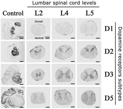
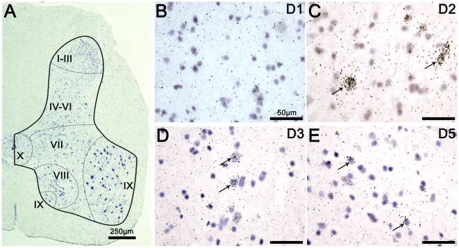
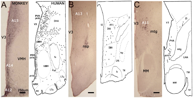
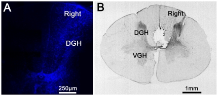
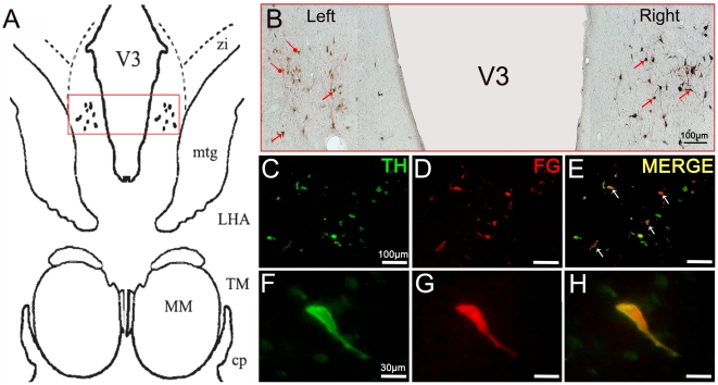
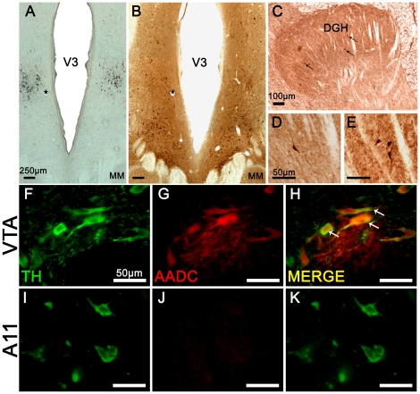
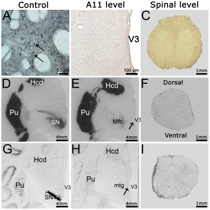
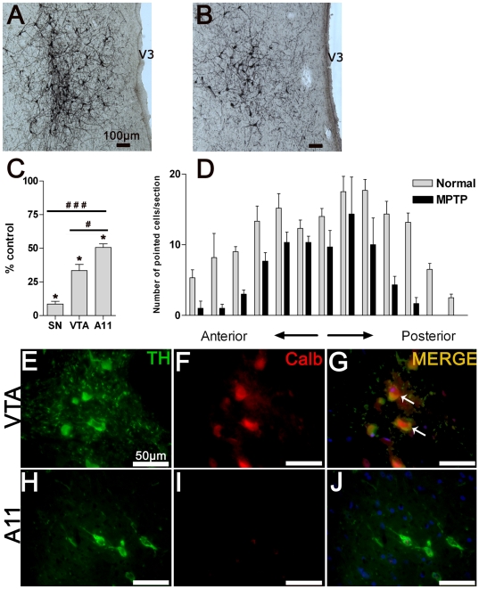
Methods and findings: In situ hybridization of spinal dopamine receptors showed that D1 receptor mRNA is absent while D2 and D5 receptor mRNAs are mainly expressed in the dorsal horn and D3 receptor mRNA in both the dorsal and ventral horns. Unilateral injections of the retrograde tracer Fluoro-Gold (FG) into the cervical spinal enlargement labeled A11 hypothalamic neurons quasi-exclusively among dopamine areas. Detailed immunohistochemical analysis suggested that these FG-labeled A11 neurons are tyrosine hydroxylase-positive but dopa-decarboxylase and dopamine transporter-negative, suggestive of a L-DOPAergic nucleus. Stereological cell count of A11 neurons revealed that this group is composed by 4002±501 neurons per side. A 1-methyl-4-phenyl-1, 2, 3, 6-tetrahydropyridine (MPTP) intoxication with subsequent development of a parkinsonian syndrome produced a 50% neuronal cell loss in the A11 group.

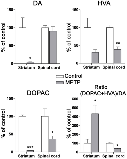
Conclusion: The diencephalic A11 area could be the major source of L-DOPA in the NHP spinal cord, where it may play a role in the modulation of sensorimotor integration through D2 and D3 receptors either directly or indirectly via dopamine formation in spinal dopa-decarboxylase-positives cells.


Conflict of interest statement

Competing Interests: HC is salaried of Cynbiose. SMC and PR are salaried employees of Motac Neuroscience Ltd. IG received research donation from GlaxoSmithKline laboratory. This does not alter the authors' adherence to all the PLoS ONE policies on sharing data and materials.

Figures

Figure 1
Figure 1. Macroscopic detection of dopamine receptors within the lumbar cord of non-human primate.
Representative film autoradiograms after radioactive in situ hybridization targeted against the mRNA DA receptors and transporter in lumbar spinal cord transverse sections at different levels (L2, L4 and L5) and in frontal brain sections (positive control). Note that D2 and D3 subtypes are the most expressed DA receptors: D2 receptors were more highly expressed in dorsal horn of the spinal cord whereas D3 receptors showed lower levels of expression with much wider distribution within the gray matter of the spinal cord. Expression of the D1 subtype was not detected whereas the D5 subtype was poorly expressed. Abbreviations: Hcd = Head of Caudate nucleus; Pu = Putamen; IClj = Islands of Calleja; Co = Cortex.
Figure 2
Figure 2. Microscopic detection of D2, D3 and D5 dopaminergic receptors in the lumbar cord.
A. Reconstructed micrograph showing a representative distribution of neurons and glial cells in a section from the lumbar spinal cord (L4). The distribution of neurons and glial cells is shown with toluidine blue staining. Surrounding white and internal gray matter are delineated with a thick black line. Division of spinal cord into laminae is approximate and separated with fine black lines according to the Rexed laminae description. Note that in laminae I-III, neuronal cell body diameters are generally the smallest. In laminae IX, neuronal cells bodies are generally the biggest (probably corresponding to motoneurons). B. Illustrative brightfield micrograph showing the detection of D1 labeling in the dorsal part of a lumbar spinal cord section. Note the lack of D1 positive cells. C. Illustrative brightfield micrograph showing the detection of D2 labeling in the dorsal part of a lumbar spinal cord section. The arrows point to typical positive cells for D2 labeling. D. Illustrative brightfield micrograph showing the detection of D3 labeling in the ventral part of a lumbar spinal cord section. The arrows point to typical positive cells for D3 labeling. E. Illustrative brightfield micrograph showing the detection of D5 labeling in the dorsal part of lumbar spinal cord section. The arrows point to typical positive cells for D5 labeling. Note that D2 labeling was mainly found in laminae I to VI, that the D3 labeling showed a wider distribution in laminae I to X and that the D5 labeling was mainly found in laminae I to III.
Figure 3
Figure 3. Characterization of tyrosine hydroxylase-positive neurons within the diencephalon in the non-human primate compared to human.
A. At the anterior hypothalamus level (AC-3 mm) where TH-IR regions A12, A14 and A15 are delineated. B. At the medial hypothalamus (AC-4 mm) where region A13 is delineated. C. At the posterior hypothalamus level (AC-5 mm) where the TH-IR A11 region is delineated. Note the remarkable concordance between NHP and human TH-IR distributions. Representative drawings of the human diencephalon are taken from Kitahama et al., 1998 with the permission of Elsevier (License N°2361961459060). Abbreviations: AC = Anterior Commissure; ARH = Arcuate Hypothalamic Nucleus; cp = Cerebral Peduncle; DHA = Dorsal Hypothalamic Area; DMH = Dorsomedial Hypothalamic Nucleus; fx = fornix; LHA = Lateral hypothalamic Area; Ltu = Lateral tuberal nucleus; nsp = nigrostriatal dopaminergic pathway; PaF = Parafornical nucleus; PEH = Periventricular Hypothalamic nucleus; PHA = Posterior Hypothalamic Area; TM = Tuberomammillary nucleus; V3 = Third Ventricle; VMH = Ventral Medial Hypothalamus; ZI = Zona Incerta.
Figure 4
Figure 4. Histological analyses of FluoroGold injections into the lumbar spinal cord.
A. Visualization of FG labeling by fluorescence microscope providing ultraviolet excitation light. B. Visualization of FG labeling by immunohistochemistry directed against FG. Note that FG was injected into the right dorsal horn of the cervical spinal cord and that there was a slight diffusion of the marker within the contralateral side. Abbreviations: DGH = Dorsal Gray Horn; VGH = Ventral Gray Horn.
Figure 5
Figure 5. Retrograde labeling of A11 neurons projecting to the spinal cord.
A. Schematic representation of the hypothalamic A11 area in which TH-IR neurons were reached by FluoroGold. The dots represent the localization of TH-IR A11 neurons. The red frame represents the area where representative micrographs were taken. B. Representative micrograph showing a reconstructed overview of the TH immunopositive and FG retrograde labeled cells in the A11 group. Immunoreactivity was revealed with Novared kit for TH neurons (Red) and with SG kit for FG neurons (Blue). Double-stained neurons (TH-FG) were labeled in a blue-red combination (i.e. black). The black arrows point to typical double-stained cells. The round heads arrows point to TH-stained neurons. Note that no single FG-stained neurons were found and that the majority of double-stained neurons are located on the ipsilateral side of spinal injections (right) C–H: Representative double-fluorescent immunostaining of TH (green) and FG (red) obtained under a confocal laser-scanning microscope in the A11 posterior hypothalamic group. The white arrows point to typical double-stained cells. Note the colocalization between the TH-positive and FG-positive neurons within the A11 region. Abbreviations: cp = Cerebral Peduncle; FG = FluoroGold; MM = Medial Mammillary nucleus; LHA = Lateral Hypothalamic Area; TH = Tyrosine Hydroxylase; TM = Tuberomammillary nucleus; V3 = Third Ventricle; ZI = Zona Incerta.
Figure 6
Figure 6. Dopamine beta-hydroxylase (DBH) expression in the locus coeruleus (A) and the posterior hypothalamus (B).
Note the lack of DBH labeling in the posterior hypothalamus (A11 area). Abbreviations: scp = superior cerebellar peduncle; V3 = Third Ventricle.
Figure 7
Figure 7. Aromatic aminoacid decarboxylase (AADC) expression in posterior hypothalamus and spinal cord.
A–B: Immunohistochemistry targeted against TH (A) and AADC (B) processed on adjacent sections of the posterior hypothalamus. Note the lack of AADC labeling in the region of TH-IR A11 neurons. Stars label the same blood vessel profiles in adjacent sections (A and B). C–E: Immunohistochemistry targeted against AADC within the dorsal horn aspect of the spinal cord. The black arrows point to typical AADC positive neurons. F–K: Double-fluorescent immunostaining of TH (green) and AADC (red) obtained under a confocal laser-scanning microscope in a section through the VTA (F–H) and A11 (I–K) groups. The white arrows point to typical double-stained cells in VTA. Note the absence of AADC-positive neurons within the A11 region. Abbreviations: AADC = Aromatic Aminoacid Decarboxylase; DGH = Dorsal Gray Horn; MM = Mammillary nucleus; TH = Tyrosine Hydroxylase; V3 = Third Ventricle.
Figure 8
Figure 8. Absence of dopamine transporter expression in the diencephalospinal pathway.
A–C: Immunohistochemistry targeted against the DAT. A. Representative micrograph of DAT labeling positive neurons in the ventral tegmental area. The arrows point to typical immunopositive neurons. B. Representative micrograph showing the absence of DAT labeling at the posterior hypothalamus level. C. Representative micrograph showing the absence of DAT labeling in a lumbar spinal section. D–F: Representative DAT binding autoradiographs in the substantia nigra (D), posterior hypothalamus (E) and lumbar spinal cord (F). Note the absence of DAT expression in the posterior hypothalamus and spinal cord, in contrast with the intense expression in the substantia nigra and striatum (positive controls). The arrow points to the approximate location of A11 area. G–I: Film autoradiograms after radioactive in situ hybridization targeting the DAT mRNAs in the substantia nigra (G), posterior hypothalamus (H) and lumbar spinal cord (I). Note the absence of DAT mRNA expression in the posterior hypothalamus and spinal cord, in contrast with the intense expression in the substantia nigra (positive control). The arrow points to the approximate location of A11 area. Abbreviations: Hcd = Head of Caudate nucleus; Mfb = Medial forebrain bundle; MM = Medial Mammillary nucleus; mtg = Mammilotegmental fasciculus; Pu = Putamen; SN = Substantia Nigra; V3 = Third Ventricle.
Figure 9
Figure 9. Effect of the neurotoxin MPTP on hypothalamic A11 neurons.
A. Representative microphotograph of TH-IR A11 neurons in a control animal. B. Representative microphotograph of TH-IR A11 neurons in an MPTP-intoxicated animal with full parkinsonism. Note the large cell loss within the A11 area. C. Mean (±SD) percentage of total number of TH-IR neurons remaining following MPTP intoxication within the substantia nigra, ventral tegmental area and A11 group (*P<0.0005, comparison between MPTP-treated animals (n = 2) and control animals (n = 4); # P<0.02 and ### P<0.0003, comparison between dopaminergic groups following MPTP intoxication, two-tailed P value, unpaired t-test). Note the difference in cell loss following MPTP intoxication between the A11 group and the various DA regions. D. TH-IR counted cells were mapped in individual sections from anterior to posterior hypothalamic A11 area with 200 µm section intervals. Note the general cell loss at different levels of the A11 area in MPTP-treated animals (n = 2) compared to controls (n = 4). E–J: Representative micrograph of TH (green) and CALB (red) double fluorescent immunostaining obtained under a confocal laser-scanning microscope in VTA (E–G) and A11 (H–J) sections. The white arrows point to typical double-stained neurons. Note the absence of CALB-positive neurons within the A11 region. Abbreviations: Calb = Calbindin 28 k; TH = Tyrosine Hydroxylase; V3 = Third Ventricle.
Figure 10
Figure 10. HPLC detection of striatal and spinal dopamine and its metabolite levels in control and MPTP-treated monkeys.
Mean (% of control ± SD) regional DA, HVA and DOPAC levels were obtained from 3 controls and 3 MPTP-treated animals. Note the significant drop in DA and DOPAC levels in the striatum following MPTP intoxication. In the lumbar spinal cord, note the absence of DA level modification, the significant decrease in metabolite levels and the DA turnover index (ratio (HVA+DOPAC)/DA) in MPTP-treated animals compared to controls (*P<0.05; **P<0.001; ***P<0.0001, two-tailed P value, unpaired t-test). Abbreviations: DA = Dopamine, DOPAC = 3,4-Dihydroxyphenylacetic acid, HVA = Homovanillic acid.

References

    1. van Os J, Kapur S. Schizophrenia. Lancet. 2009;374:635–645. - PubMed
    1. Braak H, Del Tredici K, Rub U, de Vos RA, Jansen Steur EN, et al. Staging of brain pathology related to sporadic Parkinson's disease. Neurobiol Aging. 2003;24:197–211. - PubMed
    1. Spiga S, Lintas A, Diana M. Addiction and cognitive functions. Ann N Y Acad Sci. 2008;1139:299–306. - PubMed
    1. Bjorklund A, Skagerberg G. Evidence for a major spinal cord projection from the diencephalic A11 dopamine cell group in the rat using transmitter-specific fluorescent retrograde tracing. Brain Res. 1979;177:170–175. - PubMed
    1. Hokfelt T, Phillipson O, Goldstein M. Evidence for a dopaminergic pathway in the rat descending from the A11 cell group to the spinal cord. Acta Physiol Scand. 1979;107:393–395. - PubMed

Publication types