. 2010 Oct 12;5(10):e13251.
doi: 10.1371/journal.pone.0013251.

Treating viral exacerbations of chronic obstructive pulmonary disease: insights from a mouse model of cigarette smoke and H1N1 influenza infection

Affiliations

Treating viral exacerbations of chronic obstructive pulmonary disease: insights from a mouse model of cigarette smoke and H1N1 influenza infection

Carla M T Bauer et al. PLoS One. .

Abstract

Background: Chronic obstructive pulmonary disease is a progressive lung disease that is punctuated by periods of exacerbations (worsening of symptoms) that are attributable to viral infections. While rhinoviruses are most commonly isolated viruses during episodes of exacerbation, influenza viruses have the potential to become even more problematic with the increased likelihood of an epidemic.

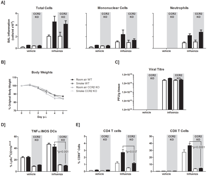
Methodology and principal findings: This study examined the impact of current and potential pharmacological targets namely the systemic corticosteroid dexamethasone and the peroxisome proliferator-activated receptor-gamma agonist pioglitazone on the outcome of infection in smoke-exposed mice. C57BL/6 mice were exposed to room air or cigarette smoke for 4 days and subsequently inoculated with an H1N1 influenza A virus. Interventions were delivered daily during the course of infection. We show that smoke-exposed mice have an exacerbated inflammatory response following infection. While smoke exposure did not compromise viral clearance, precision cut lung slices from smoke-exposed mice showed greater expression of CC (MCP-1, -3), and CXC (KC, MIP-2, GCP-2) chemokines compared to controls when stimulated with a viral mimic or influenza A virus. While dexamethasone treatment partially attenuated the inflammatory response in the broncho-alveolar lavage of smoke-exposed, virally-infected animals, viral-induced neutrophilia was steroid insensitive. In contrast to controls, dexamethasone-treated smoke-exposed influenza-infected mice had a worsened health status. Pioglitazone treatment of virally-infected smoke-exposed mice proved more efficacious than the steroid intervention. Further mechanistic evaluation revealed that a deficiency in CCR2 did not improve the inflammatory outcome in smoke-exposed, virally-infected animals.

Conclusions and significance: This animal model of cigarette smoke and H1N1 influenza infection demonstrates that smoke-exposed animals are differentially primed to respond to viral insult. While providing a platform to test pharmacological interventions, this model demonstrates that treating viral exacerbations with alternative anti-inflammatory drugs, such as PPAR-gamma agonists should be further explored since they showed greater efficacy than systemic corticosteroids.


Conflict of interest statement

Competing Interests: The work described herein was in-part funded by the Canadian Institutes of Health Research, and AstraZeneca, Sweden. JDT at AstraZeneca participated in study design and preparation of the manuscript. This does not alter the authors' adherence to all the PLoS ONE policies on sharing data and material.

Figures

Figure 1
Figure 1. Influenza infection of smoke-exposed animals.
C57BL/6 mice were exposed to room air (open bars) or cigarette smoke (closed bars). Subsequently, mice were given a PBS vehicle or inoculated with influenza A virus intranasally. (A) Broncho-alveolar lavage (BAL) samples were obtained and total cells were enumerated at the indicated times post-infection. Mononuclear cell and neutrophil differentials were also assessed. (B) Light photomicrographs of representative hematoxylin and eosin –stained cross sections of lung tissue were taken at 5 days post-infection. Data are presented as means ± SEMs for n = 3–12 animals per group.
Figure 2
Figure 2. Viral titres and type I interferon responses.
C57BL/6 mice were exposed to room air (open bars) or cigarette smoke (closed bars). Mice were infected with influenza A virus. (A) Viral titres were determined in lung homogenates at the indicated times post-infection. (B) The presence of type I interferons at day 3 post-infection was also determined by plating the indicated dilutions of BAL on confluent monolayers of IRF-3 deficient mouse embryonic fibroblasts and performing a vesicular-stomatitis virus (VSV) plaque reduction assay. 100% relative intensity indicates the absence of type I interferon activity, whereas 0% relative intensity indicates sufficient levels of type I interferon to completely prevent VSV-GFP replication. Data are presented as the means ± SEMs for n = 3–17 animals (A), and n = 5 animals (B) per group.
Figure 3
Figure 3. Expression of mediators from precision cut lung slices.
C57BL/6 mice were exposed to room air (open bars) or cigarette smoke (closed bars). (A) Precision cut lung slices were generated and cultured ex vivo. Induction of MCP-1 and MCP-3 (B) and GCP-2, KC, and MIP-2 (C) transcripts, normalized to levels of a house keeping gene, GAPDH, in the same sample and expressed relative to an untreated sample, were obtained from dsRNA (polyI:C) stimulated and influenza infected precision cut lung slices by real-time quantitative RT-PCR. Induction of ISG-15 and IRF-7 were determined similarly (D). Data are presented as means ± SEMs for n = 5–15 lung slices generated from 3 independent experiments (polyI:C studies) and a representative experiment (influenza studies).
Figure 4
Figure 4. Effect of dexamethasone in smoke-exposed influenza-infected mice.
Female C57BL/6 mice were exposed to room air (open bars) or cigarette smoke (closed bars). One hour prior to treatment with vehicle or inoculation with influenza A virus, mice were gavaged with 3mg/kg of dexamethasone (dex). Mice were gavaged daily with 3 mg/kg of dex. (A) BAL samples were obtained at 5 days post-infection and total cells were enumerated. Mononuclear cell and neutrophil differentials were also assessed. Clinical presentation in animals at the time of sacrifice (B), and viral titres (C) were also determined. Clinical presentation (D), and viral titres (E) were also assessed in mice sacrificed at day 7 post-infection. Data are presented as the means ± SEMs for n = 4–6 animals per group.
Figure 5
Figure 5. Effect of a PPAR-γ agonist, pioglitazone, on antiviral responses in smoke-exposed mice.
C57BL/6 mice were exposed to room air (open bars) or cigarette smoke (closed bars). Starting on the second day of smoke exposure, mice were gavaged daily with 60 mg/kg of pioglitazone (Pio). Following four days of smoke-exposure, mice were given a sterile vehicle or inoculated with influenza A virus intranasally. (A) Broncho-alveolar lavage (BAL) samples were obtained at 5 days post-infection and total cells were enumerated. Mononuclear cell and neutrophil differentials were also assessed. (B) Body weight was monitored throughout the course of the viral infection and expressed relative to body weight on the day 0 of infection. (C) Viral titres were also determined. Percentages of TNFα/iNOS producing dendritic cells (D), and activated CD4+ and CD8+ T cells (E) were obtained from the live gate of CD45+ cells. Data are presented as the means ± SEMs for n = 5–6 animals per group.
Figure 6
Figure 6. Effect of a CCR2 deficiency on antiviral responses in smoke-exposed mice.
C57BL/6 wild-type (WT) or CCR2 knock-out (KO) mice were exposed to room air (open bars) or cigarette smoke (closed bars). Subsequently, mice were given a sterile vehicle or inoculated with influenza A virus intranasally. (A) Broncho-alveolar lavage (BAL) samples were obtained at 5 days post-infection and total cells were enumerated. Mononuclear cell and neutrophil differentials were also assessed. Percent body weight change throughout the viral infection was documented (B), and viral titres were also determined (C). Percentages of TNFα/iNOS producing dendritic cells (D), and activated CD4+ and CD8+ T cells (E) were obtained from the live gate of CD45+ cells. Data are presented as the means ± SEMs for n = 5 animals per group.

References

    1. Rodriguez-Roisin R. Toward a consensus definition for COPD exacerbations. Chest. 2000;117:398S–401S. - PubMed
    1. Wesseling G. Occasional review: influenza in COPD: pathogenesis, prevention, and treatment. Int J Chron Obstruct Pulmon Dis. 2007;2:5–10. - PMC - PubMed
    1. Mannino DM, Buist AS. Global burden of COPD: risk factors, prevalence, and future trends. Lancet. 2007;370:765–773. - PubMed
    1. Wedzicha JA. Airway infection accelerates decline of lung function in chronic obstructive pulmonary disease. Am J Respir Crit Care Med. 2001;164:1757–1758. - PubMed
    1. Mallia P, Johnston SL. Mechanisms and experimental models of chronic obstructive pulmonary disease exacerbations. Proc Am Thorac Soc. 2005;2:361–366; discussion 371–362. - PubMed

Publication types

MeSH terms