. 2010 Oct 12;5(10):e12947.
doi: 10.1371/journal.pone.0012947.

Catalytic mechanism of cellulose degradation by a cellobiohydrolase, CelS

Affiliations

Catalytic mechanism of cellulose degradation by a cellobiohydrolase, CelS

Moumita Saharay et al. PLoS One. .

Abstract

The hydrolysis of cellulose is the bottleneck in cellulosic ethanol production. The cellobiohydrolase CelS from Clostridium thermocellum catalyzes the hydrolysis of cello-oligosaccharides via inversion of the anomeric carbon. Here, to examine key features of the CelS-catalyzed reaction, QM/MM (SCCDFTB/MM) simulations are performed. The calculated free energy profile for the reaction possesses a 19 kcal/mol barrier. The results confirm the role of active site residue Glu87 as the general acid catalyst in the cleavage reaction and show that Asp255 may act as the general base. A feasible position in the reactant state of the water molecule responsible for nucleophilic attack is identified. Sugar ring distortion as the reaction progresses is quantified. The results provide a computational approach that may complement the experimental design of more efficient enzymes for biofuel production.


Conflict of interest statement

Competing Interests: The authors have declared that no competing interests exist.

Figures

Figure 1
Figure 1. Model of cellobiohydrolase CelS structure in complex with celloheptaose (green), and cellobiose, colored in white (hydrogen), red (oxygen), and cyan (carbon).
The sugar unit at subsite −1 was modelled manually. The substrate, celloheptaose, spans between subsites −1 to −7 in the substrate-binding tunnel, and cellobiose is bound in the cleft region between subsites +1 and +2. This figure was made using the VMD software .
Figure 2
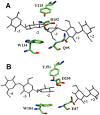
Figure 2. Structural comparison of active sites of (A) family 8 CelA and (B) family 48 CelS.
Substrate is shown in Gray.
Figure 3
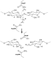
Figure 3. Schematic representation of inverting reaction mechanism in CelS for hydrolysis of glycosidic bond C1-O4.
The catalytic residues Glu87, Asp255, and nucleophilic water molecule (W1) are shown. Anomeric carbon atom at subsite −1 and leaving group oxygen atom at subsite +1 are C1 and O4, respectively. The thin arrows represent electron transfer between atoms. Distances between atoms are shown in red.
Figure 4
Figure 4. Potential of mean force for hydrolysis reaction.
Figure 5
Figure 5. Snapshot of reactant state from QM/MM molecular dynamics free energy trajectories.
Only the active site residues, glucosyl units −1 and +1, acid catalyst Glu87, base catalyst Asp255, nucleophilic water, and a second water molecule shown in gold (omitted Hydrogen atoms for clarity), are shown here. All backbone atoms of Glu87 and Asp255 are omitted for clarity. Important distances are shown by black lines.
Figure 6
Figure 6. Snapshot of transition state from QM/MM molecular dynamics free energy trajectories.
Other figure specifications are similar to Fig 5.
Figure 7
Figure 7. Snapshot of product state from QM/MM molecular dynamics free energy trajectories.
Other figure specifications are similar to Fig 5.
Figure 8
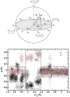
Figure 8. Upper panel: Puckering coordinates (Q,,) for six membered ring.
Lower panel: Projection of puckering coordinates (qformula image, and qformula image) sampled by molecular dynamics trajectories along the reaction coordinate rc2. qformula image and qformula image values are shown in gray and black, respectively. Regions corresponding to reactant (R), around transition state (TS), and product (R) are highlighted by boxes.
Figure 9
Figure 9. Stoddart diagram showing ring conformations at subsite −1 with respect to q and q.
Conformations for reactant (brown circle), transition (red triangle), and product (blue sqare) states are shown. Different regions are separated by dashed black lines. The central region represents undistorted and stable formula imageCformula image conformation.
Figure 10
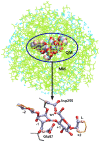
Figure 10. Setup for enzyme reaction in CelS with QM/MM method.
QM region (VDW representation) consists of catalytic residues (Asp255 and Glu87), nucleophilic water (W1), and active part of substrate (subsites −1, and +1), while rest of enzyme (green), substrate (orange), and water (cyan) are in MM region. Inset shows only QM region and hydrogen link atoms (pink) used as boundary between MM and QM.

References

    1. Sticklen M. Plant genetic engineering for biofuel production: towards affordable cellulosic ethanol. Nat Rev Gent. 2008;9:433–443. - PubMed
    1. Himmel M, Ding S, Johnson D, Adney W, Nimlos M, et al. Biomass recalcitrance: Engineering plants and enzymes for biofuels production. Science. 2007;315:804. - PubMed
    1. Demain A, Newcomb M, Wu J. Cellulase, Clostridia, and Ethanol. Microbiol Mol Biol Rev. 2005;69:124. - PMC - PubMed
    1. Beguin P, Aubert J. The biological degradation of cellulose. FEMS Microbiol Rev. 1994;13:25–58. - PubMed
    1. Creuzet N, Berenger J, Frixon C. Characterization of exoglucanase and synergistic hydrolysis of cellulose in Clostridium stercorarium. FEMS Microbiol Lett. 1983;20:347–350.

Publication types

MeSH terms

Substances

LinkOut - more resources