. 2011 Feb 7;136(3):473-8.
doi: 10.1039/c0an00609b. Epub 2010 Oct 21.

High-throughput 3D spheroid culture and drug testing using a 384 hanging drop array

Affiliations

High-throughput 3D spheroid culture and drug testing using a 384 hanging drop array

Yi-Chung Tung et al. Analyst. .

Abstract

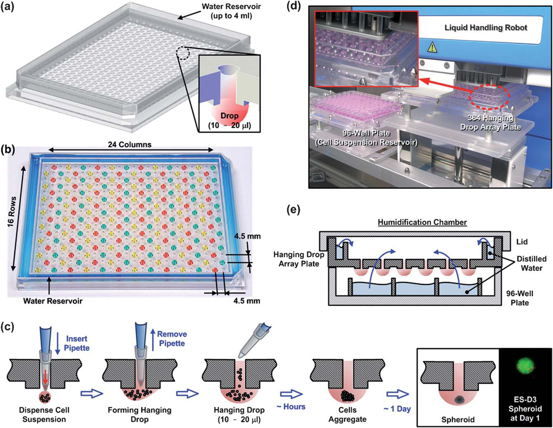
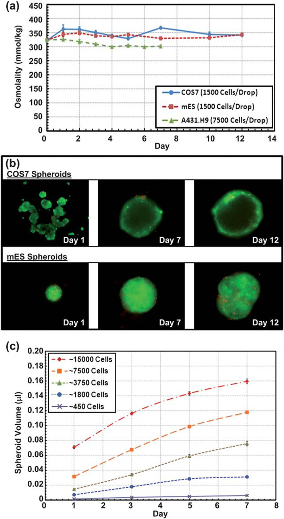
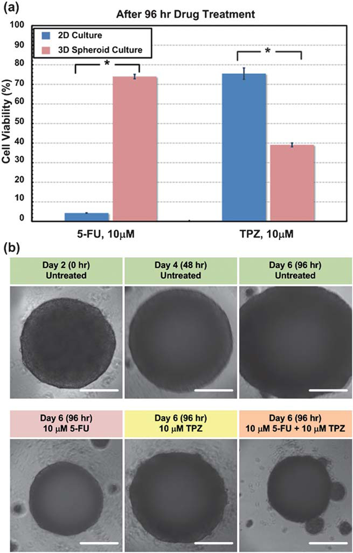
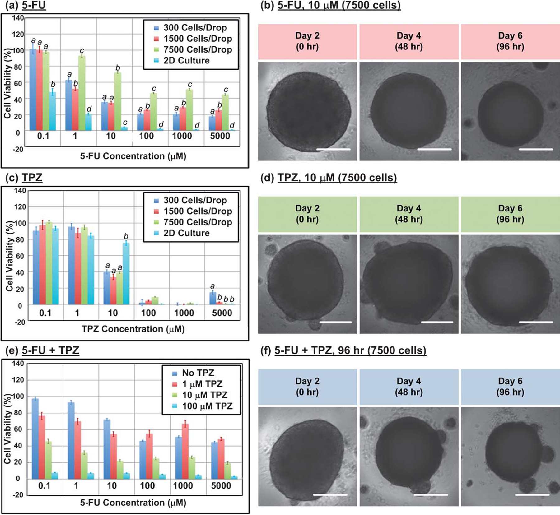
Culture of cells as three-dimensional (3D) aggregates can enhance in vitro tests for basic biological research as well as for therapeutics development. Such 3D culture models, however, are often more complicated, cumbersome, and expensive than two-dimensional (2D) cultures. This paper describes a 384-well format hanging drop culture plate that makes spheroid formation, culture, and subsequent drug testing on the obtained 3D cellular constructs as straightforward to perform and adapt to existing high-throughput screening (HTS) instruments as conventional 2D cultures. Using this platform, we show that drugs with different modes of action produce distinct responses in the physiological 3D cell spheroids compared to conventional 2D cell monolayers. Specifically, the anticancer drug 5-fluorouracil (5-FU) has higher anti-proliferative effects on 2D cultures whereas the hypoxia activated drug commonly referred to as tirapazamine (TPZ) are more effective against 3D cultures. The multiplexed 3D hanging drop culture and testing plate provides an efficient way to obtain biological insights that are often lost in 2D platforms.


Figures

Fig. 1
Fig. 1
(a) Illustration of the designed 384 hanging drop spheroid culture array plate, and its cross-sectional view. (b) Photo and key dimensions of the array plate. (c) Cartoon of the hanging drop formation process in the array plate. The pipette tip is first inserted through the access hole to the bottom surface of the plate, and cell suspension is subsequently dispensed. Cell suspension is quickly attracted to the hydrophilic plate surface and a hanging drop is quickly formed and confined within the plateau. Within hours, individual cells start to aggregate and eventually form into a single spheroid around 1 day. (d) Photo of the 384 hanging drop array plate operated with liquid handling robot capable of simultaneously pipetting 96 cell culture sites. (e) Cartoon of the final humidification chamber used to culture 3D spheroids in the hanging drop array plate. The 384 hanging drop array plate is sandwiched between a 96-well plate filled with distilled water and a standard-sized plate lid. Distilled water from the bottom 96-well plate and the peripheral water reservoir prevent serious evaporation of the small volume hanging drops.
Fig. 2
Fig. 2
(a) Osmolality of COS7, mES, and A431.H9 cell spheroids with various cell populations over a 7- and 12-day culture. Data are expressed as the mean s.e.m. (b) Fluorescence images of live/dead stained COS7 and mES cell spheroids over a 12-day culture. (c) Volume of A431.H9 spheroids over a 7-day culture for various initial cell numbers per spheroid. n = 14 for each initial cell number condition. Data are expressed as the mean ± s.e.m.
Fig. 3
Fig. 3
(a) Bar graph of the cell viability at 10 μM 5-FU, and 10 μM TPZ 96 h after drug treatment for 2D A431.H9 monolayer culture and 7500-cell A431.H9 3D spheroid culture conditions. For both drugs, the viability of A431.H9 cells was statistically different between 2D monolayer and 3D spheroid culture conditions. Statistical significance is determined by two-tailed Student’s t-Test (*, P < 0.01) P = 1.75 × 10⁻¹⁶ for 5-FU, P = 1.22 × 10⁻⁶ for TPZ. n = 8 for 2D culture condition and Data are expressed as the n = 14 for 3D spheroid culture condition. Mean × s.e.m. (b) Time-lapse images of control untreated 7500-cell A431.H9 spheroid, and spheroids treated with 10 μM 5-FU, 10 μM TPZ, and 10 μM 5-FU + 10 μM TPZ 96 h after treatment. Scale bar is 200 μm.
Fig. 4
Fig. 4
(a) Bar graph of the cell viability at various 5-FU concentrations 96 h after drug treatment for 300, 1500, and 7500-cell A431.H9 spheroids and 2D culture condition. Different letters between culture conditions (spheroid size or 2D) within a 5-FU concentration represent a significant difference between the spheroid sizes or 2D (a, b, c, d = p < 0.01). (b) Time-lapse images of 7500-cell A431.H9 spheroids treated with 10 μM 5-FU. (c) Bar graph of the cell viability at various TPZ concentrations 96 h after drug treatment for 300, 1500, and 7500-cell A431.H9 spheroids and 2D culture condition. Different letters between culture conditions (spheroid size or 2D) within a TPZ concentration represent a significant difference between the spheroid sizes or 2D (a, b = p < 0.01). (d) Time-lapse images of 7500-cell A431.H9 spheroids treated with 10 μM TPZ. (e) Bar graph of the cell viability at various 5-FU concentrations 96 h after drug treatment for 7500-cell A431.H9 spheroids with 0, 1, 10, and 100 μM TPZ. (f) Time-lapse images of 7500-cell A431.H9 spheroids treated with 10 μM 5-FU + 10 μM TPZ. Statistical analysis was performed by ANOVA followed by Holm-Sidak tests. Spheroid size or 2D groups that are statistically significantly different are designtated with different letters (a, b, c, d). n = 8 for 2D culture condition and n 14 for 3D spheroid culture condition. Data are expressed as the mean ± s.e.m. Scale bar is 200 μm.

References

    1. Pampaloni F, Reynaud EG and Stelzer EHK, Nat. Rev. Mol. Cell Biol, 2007, 8, 839–845. - PubMed
    1. Friedrich J, Seidel C, Ebner R and Kunz-Schughart LA, Nat. Protoc, 2009, 4, 309–324. - PubMed
    1. Kunz-Schughart LA, Freyer JP, Hofstaedter F and Ebner R, J. Biomol. Screening, 2004, 9, 273–285. - PubMed
    1. Kubbies AIM, Biomol J. Screening, 2006, 11, 922–932. - PubMed
    1. Del Duca D, Werbowetski T and Del Maestro RF, J. Neuro-Oncol, 2004, 67, 295–303. - PubMed

Publication types

MeSH terms

Substances