. 2011 Jun;68(11):1941-52.
doi: 10.1007/s00018-010-0540-2. Epub 2010 Oct 22.

Signalling pathways involved in ribonuclease-7 expression

Affiliations

Signalling pathways involved in ribonuclease-7 expression

Imran Mohammed et al. Cell Mol Life Sci. 2011 Jun.

Abstract

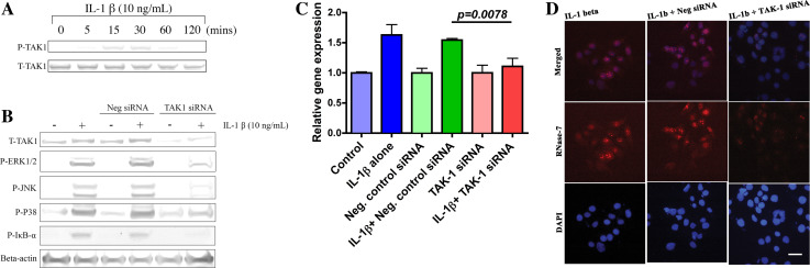
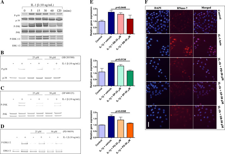
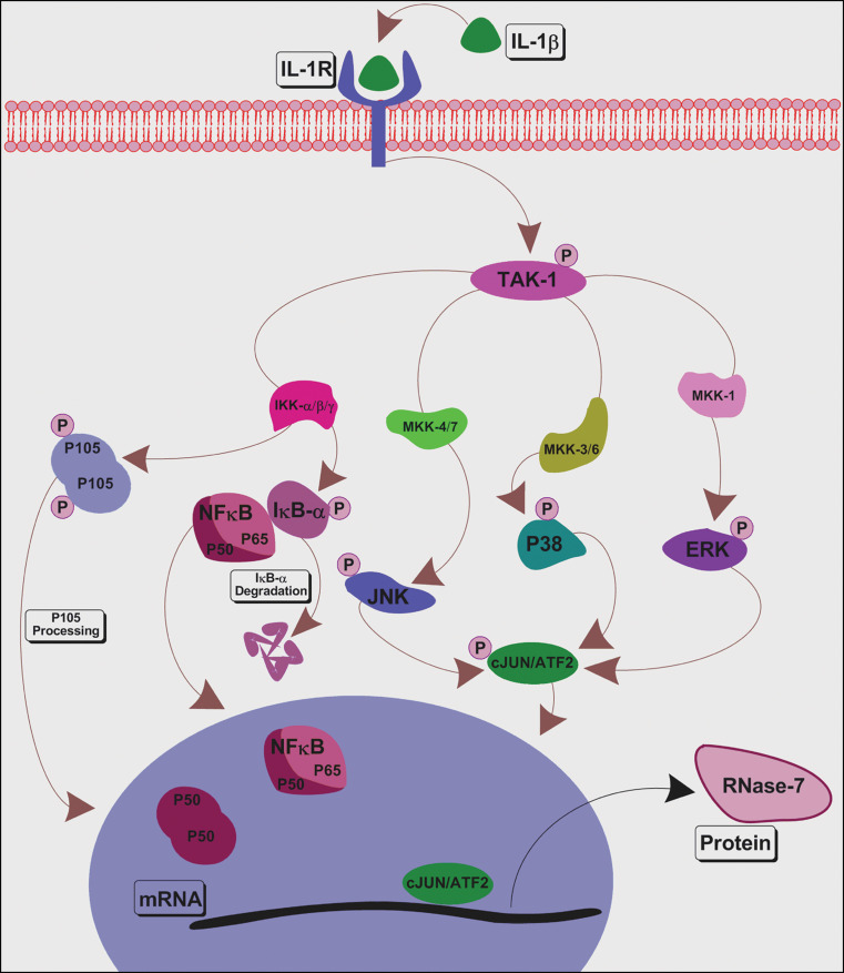
Antimicrobial peptides are host defence molecules that play a potential role in preventing infection at the epithelial surfaces. Ribonuclease (RNase)-7 has been shown to possess a broad spectrum of microbicidal activity against various pathogens. Here, we demonstrate that RNase-7 protein is localised to the superficial layers of ocular surface cells and increased in response to interleukin (IL)-1β, suggesting an active role during inflammation related to ocular surface infection. Signal transduction pathways involved in RNase-7 expression are unknown. Involvement of transforming growth factor β-activated kinase-1 (TAK-1) activated nuclear factor kappa B (NF-κB) and mitogen-activated protein kinase (MAPK) pathway molecules [c-Jun N-terminal kinase (JNK), extracellular signal-regulated kinase (ERK) and p38] were studied because of their importance in infection and inflammation. Blocking the MAPKs resulted in inhibition of RNase-7 expression in response to IL-1β. However, RNase-7 induction by IL-1β was not affected by inhibiting the NF-κB signalling pathway. In conclusion, our results indicate that RNase-7 expression is specifically mediated via MAPKs but not NF-κB signalling pathways.


Figures

Fig. 1
Fig. 1
In-vitro expression of RNase-7 in human ocular surface. a Localisation of RNase-7 protein at OS. The upper panel displays merged images of both RNase-7 (red) and nuclei (DAPI blue) staining. Negative control staining showed no immunoreactive staining. Scale bar: 100 μM. b–d Effect of IL-1β on RNase-7 expression. b Cells were incubated with IL-1β for indicated time points, and qPCR analysis was performed. c Cells were treated with different concentrations of IL-1β for 3 h. RNase-7 mRNA levels were analysed by qPCR. Data represent means ± SEM of triplicate samples repeated three times. p < 0.05. d Cells were incubated for indicated times with (+) or without (−) IL-1β and were then stained for RNase-7. Scale bar: 100 μM. All data are representative of three independent experiments
Fig. 2
Fig. 2
Transforming growth factor β-activated kinase-1 plays a central role in modulation of RNase-7 expression. a Rapid induction of TAK-1 by IL-1β. Cells treated with IL-1β for indicated time points and then cell lysates were prepared and analysed by WB. b TAK-1-dependent regulation of NF-κB and MAPKs in response to IL-1β. Cells transfected without (−) or with (+) TAK-1 siRNA or negative control siRNA were incubated with IL-1β and were then analysed with indicated Abs. c, d Knockdown of TAK-1 suppresses IL-1β induced RNase-7 expression. c Role of IL-1β/TAK-1 axis in RNase-7 mRNA expression. Following transfection without or with indicated siRNA, hCECs were incubated in the presence or absence of IL-1β for 3 h. RNase-7 mRNA was analysed by qPCR and presented as mean value ± SEM; n  =  3. The p value corresponds to the significance relative to the negative control. d Reduction in IL-1β induced RNase-7 protein levels in TAK-1 siRNA transfected hCECs. Cells transfected without or with indicated siRNA prior to incubation with IL-1β for 24 h were stained with RNase-7 antibody (red middle panel) and DAPI (blue lower panel). Scale bar 100 μM
Fig. 3
Fig. 3
RNase-7 expression is independent of NF-κB signalling pathway in hCECs. a Activation of NF-κB signalling by IL-1β. Cells were treated with IL-1β for the indicated times. The specified fractions were analysed using WB. b, c Suppression of IL-1β activated NF-κB pathway. b NAI and SC-514 reduce IL-1β-induced IκB-α phosphorylation. Cytosolic extract was prepared and analysed using WB. c Reduced nuclear translocation of NF-κB/p65 in IL-1β-treated hCECs after blocking. Nuclear lysates were prepared as indicated in SI and then analysed using WB. d, e Suppression of NF-κB does not effect IL-1β induced RNase-7 expression. d NAI and SC-514 do not reduce RNase-7 mRNA expression in IL-1β-treated hCECs. Cells pretreated without or with indicated inhibitors at various concentrations were incubated in the presence or absence of IL-1β. RNase-7 mRNA levels were measured by qPCR. e IL-1β induced RNase-7 protein levels are not altered by NF-κB pathway inhibitors. IF staining of RNase-7 (middle panel) in inhibitor-pretreated hCECs compared to those treated with IL-1β alone. Scale bar: 100 μM. f, g Knockdown of NF-κB1/p105 does not alter IL-1β induced RNase-7 levels. f NF-κB1/p105 siRNA does not affect RNase-7 mRNA levels. Cells transfected without or with indicated siRNA prior to incubation in the presence or absence of IL-1β were then analysed by qPCR for RNase-7 mRNA levels. The p value corresponds to the significance relative to the negative control. g Unchanged RNase-7 protein levels in NF-κB1/p105 siRNA transfected hCECs. Cells transfected without or with indicated siRNA prior to incubation with IL-1β were stained for RNase-7 (middle panel) and DAPI (lower panel). Scale bar 100 μM. All data represent means ± SEM of three samples repeated three times. p < 0.05
Fig. 4
Fig. 4
Mitogen-activated protein kinases are involved in RNase-7 mRNA expression. a Activation of MAPKs. Whole cell extract was prepared and then analysed using WB. b–d Effect of inhibitors on IL-1β-induced activation of p38, JNK and ERK. Whole cell extract was prepared post-treatment without or with indicated concentrations of SB203580. b SP600125. c PD98059 and d each prior to IL-1β exposure. Western blotting analysis was performed using indicated Abs. e, f Role of MAPKs in IL-1β-induced RNase-7 expression. e Blocking MAPKs reduces IL-1β induced RNase-7 mRNA levels. Cells are pretreated with inhibitors of p38 (top), JNK (middle) and ERK (bottom) at indicated concentrations and then were incubated in absence or presence of IL-1β. RNase-7 mRNA levels were measured by qPCR. Data are presented as mean ± SEM from three independent experiments with triplicate samples. p < 0.05. f Inhibitors of MAPKs reduce IL-1β-induced RNase-7 protein levels. Immunofluorescence staining was performed post-treatment without or with specified inhibitors at indicated concentration and then were incubated in absence or presence of IL-1β for 3 h. Scale bar 100 μM. All data are representative of three independent experiments
Fig. 5
Fig. 5
Essential role of c-Jun and ATF2 in RNase-7 expression. a IL-1β activates both c-Jun and ATF2. Cell lysate was prepared as described in SI and analysed using WB. b IL-1β/MAPKs mediate activation of c-Jun and ATF2. Whole cell extract prepared following treatment without or with indicated inhibitors each prior to IL-1β exposure was analysed using WB. c–e Role of c-Jun/ATF2 in RNase-7 expression. c, d Silencing c-Jun and ATF-2 reduces IL-1β induced RNase-7 mRNA expression. Following transfection without or with negative control or c-Jun c or ATF2 siRNAs d hCECs were incubated in the presence or absence of IL-1β for 3 h. RNase-7 mRNA levels were analysed by qPCR and presented as mean value ± SEM; n = 3. The p value was calculated relative to the negative control. e Reduction in IL-1β induced RNase-7 protein levels after silencing c-Jun and ATF2. Cells transfected without or with indicated siRNAs prior to incubation with IL-1β were stained for RNase-7 (Red middle panel) and DAPI (Blue lower panel). Scale bar 100 μM. All the data are representative of three independent experiments
Fig. 6
Fig. 6
Schematic diagram representing the signalling mechanism required for induction of RNase-7 by IL-1β in hCECs. Binding of IL-1β to membrane receptor IL-1R activates a cascade of signalling events including activation of TAK-1, NF-κB, p38, JNK and ERK. Further downstream, activation and nuclear translocation of transcription factors takes place, eventually leading to transcription of RNase-7. The results obtained in this study using siRNA or pharmacological inhibitors indicate that IL-1β induced RNase-7 expression is mediated via TAK-1-dependent MAPKs, but not NF-κB pathways

Similar articles

Cited by

References

    1. Haynes RJ, Tighe PJ, Dua HS. Innate defence of the eye by antimicrobial defensin peptides. Lancet. 1998;352(9126):451–452. doi: 10.1016/S0140-6736(05)79185-6. - DOI - PubMed
    1. Haynes RJ, Tighe PJ, Dua HS. Antimicrobial defensin peptides of the human ocular surface. Br J Ophthalmol. 1999;83(6):737–741. doi: 10.1136/bjo.83.6.737. - DOI - PMC - PubMed
    1. Yount NY, Yeaman MR. Multidimensional signatures in antimicrobial peptides. Proc Natl Acad Sci USA. 2004;101(19):7363–7368. doi: 10.1073/pnas.0401567101. - DOI - PMC - PubMed
    1. Verma C, Seebah S, Low SM, Zhou L, Liu SP, Li J, Beuerman RW. Defensins: antimicrobial peptides for therapeutic development. Biotechnol J. 2007;2(11):1353–1359. doi: 10.1002/biot.200700148. - DOI - PubMed
    1. Rosenberg HF (2008) RNase A ribonucleases and host defense: an evolving story. J Leukoc Biol - PMC - PubMed

Publication types

MeSH terms