. 2011 Feb;10(1):66-79.
doi: 10.1111/j.1474-9726.2010.00646.x.

Age-related changes in mesenchymal stem cells derived from rhesus macaque bone marrow

Affiliations

Age-related changes in mesenchymal stem cells derived from rhesus macaque bone marrow

Ji Min Yu et al. Aging Cell. 2011 Feb.

Abstract

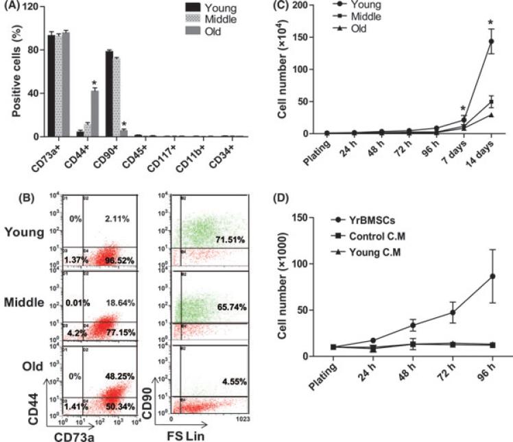
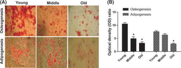
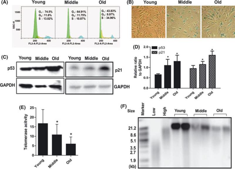
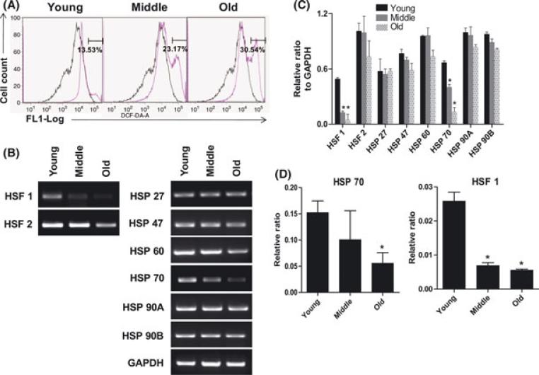
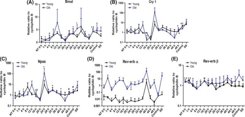
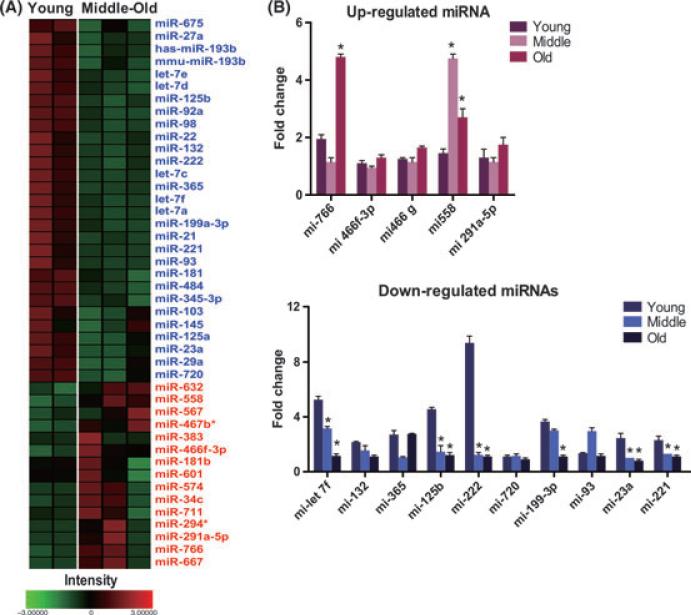
The regeneration potential of mesenchymal stem cells (MSCs) diminishes with advanced age and this diminished potential is associated with changes in cellular functions. This study compared MSCs isolated from the bone marrow of rhesus monkeys (rBMSCs) in three age groups: young (< 5 years), middle (8-10 years), and old (> 12 years). The effects of aging on stem cell properties and indicators of stem cell fitness such as proliferation, differentiation, circadian rhythms, stress response proteins, miRNA expression, and global histone modifications in rBMSCs were analyzed. rBMSCs demonstrated decreased capacities for proliferation and differentiation as a function of age. The production of heat shock protein 70 (HSP70) and heat shock factor 1 (HSF1) were also reduced with increasing age. The level of a core circadian protein, Rev-erb α, was significantly increased in rBMSCs from old animals. Furthermore, analysis of miRNA expression profiles revealed an up-regulation of mir-766 and mir-558 and a down-regulation of mir-let-7f, mir-125b, mir-222, mir-199-3p, mir-23a, and mir-221 in old rBMSCs compare to young rBMSCs. However, there were no significant age-related changes in the global histone modification profiles of the four histone core proteins: H2A, H2B, H3, and H4 on rBMSCs. These changes represent novel insights into the aging process and could have implications regarding the potential for autologous stem cells therapy in older patients.


Figures

Fig. 1
Fig. 1
Age-related changes in immunophenotype and growth curves. (A) Age-related changes in the proportion of rBMSCs expressing specific surface marker (*P < 0.05, significant difference from young rBMSCs). (B) Immunophenotypic characterization of rBMSCs in three age groups. Dot plot graphs are representative of three samples per/ each group. (C) Growth curves of rBMSCs derived from young, middle, and old age groups. The young rBMSCs expanded more rapidly than old rBMSCs. The difference in growth rate among the three groups is statistically significant (*P < 0.05, n = 4/ group). (D) Effect of CM obtained from young rBMSCs on cell growth of aged rBMSCs. Old rBMSCs were plated on 12 well plates and cultured in CM obtained from young rBMSCs. Old rBMSCs were counted every 24 h for 4 days. Data are expressed as the mean ± SED, n = 3.
Fig. 2
Fig. 2
Age-related decline in differentiation potential of rBMSCs. (A) Representative image showing Alizarin Red stained mineral deposits (top) and Oil Red O stained lipid inclusions (bottom) on cultured rBMSC for each age group. Young rBMSCs show a markedly increased level of differentiation potential at passage 2. 10× magnification. (B) Graphs represent the ratio of normalized OD of differentiated cells.
Fig. 3
Fig. 3
Increased cellular senescence in aged rBMSCs. (A) The cell cycle distribution of rBMSCs was determined by flow cytometry analysis after the cellular DNA was stained with propidium iodide (PI). The histograms of DNA content indicate the percentages of cells in G0-G1, S, and G2-M phases of the cell cycle. A representative result is shown. (B) Images of β-galactosidase staining (blue) of rBMSCs in three age groups. (C) Age-related increase in p53 and p21 protein expression was detected by Western blotting. (D) Relative ratio to GAPDH (*P < 0.05, significant difference from young rBMSCs). (E) Telomerase enzyme activity. Telomerase activity was detected by the TRAP assay. Analysis of telomerase activity showed reduced activity in old rBMSCs (*P < 0.01, significant difference from young rBMSCs). Data represent mean ± SEM of three different experiments. (F) Telomere length was determined as a mean terminal restriction fragment (TRF) length of genomic DNA based on Southern hybridization with a telomeric probe. Terminal restriction fragments were visualized, using a labeled (TTAGGG) probe.
Fig. 4
Fig. 4
Age related changes of stress responses genes. (A) ROS accumulation in rBMSCs of three age groups. DCFH-DA fluorescence of cells was determined by FACS cytometry as an indicator of ROS accumulation within the cells. A representative result is shown. Median of fluorescence intensity of unstained rBMSCs (black) and DCFH-DA stained (pink) rBMSCs of three age groups. The X axis represents log F1 fluorescence intensity; the Y axis represents cell number. (B) Alteration of stress response genes with age. Total RNA was isolated from three age groups and analyzed by RT-PCR for the indicated factor (HSF-1, HSF-2, HSP27, HSP 47, HSP 60, HSP 70, HSP 90A, and HSP 90B). A representative result is shown. (C) The quantification of stress response genes expression level via RTPCR was determined using an image analyzer. Values are mean ± SEM. *p < 0.05, significant difference compare to young rBMSCs. (D) To confirm expression of HSF 70 and HSF 1, RNA was quantified using qPCR. The graph is shown a quantitative analysis of relative expression of mRNAs at three age groups. The values were normalized using β-actin as a control. *p < 0.05, significantly difference compare to young rBMSCs.
Fig. 5
Fig. 5
The variations in relative mRNA expression for core circadian oscillator proteins: (A) Bmal1, (B) Cry1, (C) Npas, and (D) Rev-erb α, and (E) Rev-erb β in rBMSCs isolated from young and old group animals. Samples were collected every 4 h for a total of 48 h in young (black) and old (blue) rBMSCs. Cyclophilin B is used as a control. Values are mean ± SEM. (# P < 0.05 in young age group, *P < 0.05 in old age group, n = 3/ group).
Fig. 6
Fig. 6
MicroRNA expression in rBMSCs. (A) Heat map visualization and clustering of miRNA expression data. Hierarchical clustering of age dependent expression profiles among 556 miRNA in young, middle, and old rBMSCs. Relative expression levels at three age groups visualized in using a heatmap in which red indicates up-regulation in each group, while blue corresponds to down-regulation. See scale bar at bottom. (B) The fold changes in expression of the miRNAs were performed by qPCR. *P < 0.05, significant differences compare to young rBMSCs.

References

    1. Armanios M, Alder JK, Parry EM, Karim B, Strong MA, Greider CW. Short telomeres are sufficient to cause the degenerative defects associated with aging. Am. J. Hum. Genet. 2009;85:823–832. - PMC - PubMed
    1. Auricchio A, Rivera VM, Clackson T, O'Connor EE, Maguire AM, Tolentino MJ, Bennett J, Wilson JM. Pharmacological regulation of protein expression from adeno-associated viral vectors in the eye. Mol. Ther. 2002;6:238–242. - PubMed
    1. Baxter MA, Wynn RF, Jowitt SN, Wraith JE, Fairbairn LJ, Bellantuono I. Study of telomere length reveals rapid aging of human marrow stromal cells following in vitro expansion. Stem Cells. 2004;22:675–682. - PubMed
    1. Beausejour C. Bone marrow-derived cells: the influence of aging and cellular senescence. Handb. Exp. Pharmacol. 2007;180:67–88. - PubMed
    1. Bellows CG, Pei W, Jia Y, Heersche JN. Proliferation, differentiation and self-renewal of osteoprogenitors in vertebral cell populations from aged and young female rats. Mech. Ageing Dev. 2003;124:747–757. - PubMed

Publication types

MeSH terms