. 2011 Jan;201(1):97-107.
doi: 10.1111/j.1748-1716.2010.02207.x. Epub 2010 Nov 12.

Expression of acid-sensing ion channels in intestinal epithelial cells and their role in the regulation of duodenal mucosal bicarbonate secretion

Affiliations

Expression of acid-sensing ion channels in intestinal epithelial cells and their role in the regulation of duodenal mucosal bicarbonate secretion

X Dong et al. Acta Physiol (Oxf). 2011 Jan.

Abstract

Aims: As little is currently known about acid-sensing ion channels (ASICs) in intestinal epithelial cells, the aims of the present study were to investigate the expression and function of ASICs in intestinal epithelial cells, particularly their physiological role in the acid-stimulated duodenal mucosal bicarbonate secretion (DMBS).

Methods: RT-PCR and digital Ca²(+) imaging were used to determine the expression and function of ASICs in HT29 cells and SCBN cells, intestinal epithelial crypt cell lines. The acid-stimulated DMBS was measured in C57 black mice in vivo to study the role of ASICs in this physiological process.

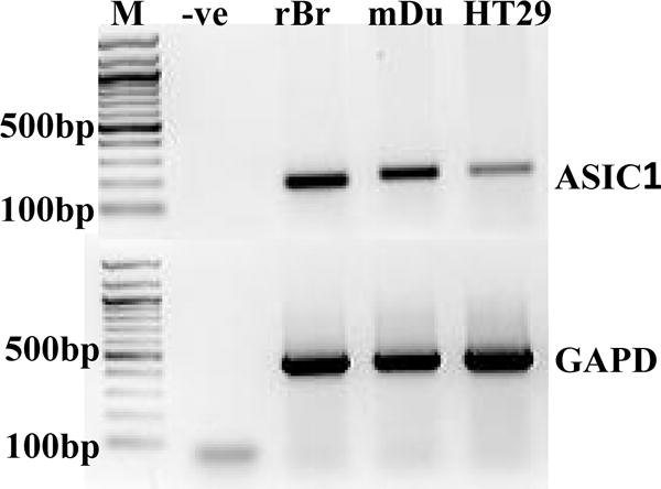
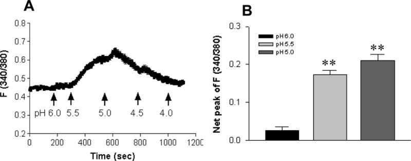
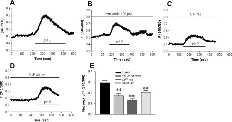
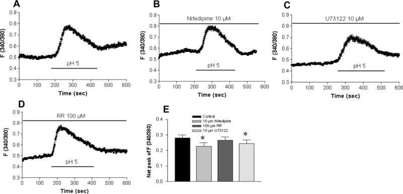
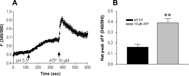
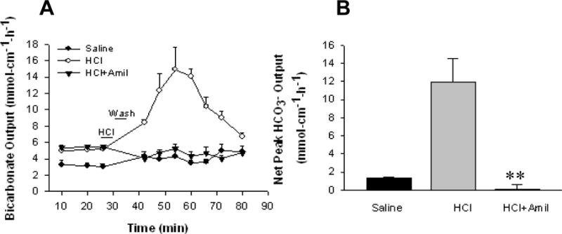
Results: ASIC1a mRNA expression was detected in the duodenal mucosa stripped from mice and epithelial cell lines, in which cytoplasmic free Ca²(+) ([Ca²(+) ](cyt)) in response to extracellular acidosis was also increased. In Ca²(+) -containing solutions, acidosis (pH 6.0-5.0) raised [Ca²(+) ](cyt) in both HT29 cells and SCBN cells in a similar pH-dependent manner. Acidosis-induced increase in [Ca²(+) ](cyt) was markedly inhibited by amiloride (an ASICs blocker), SK&F96365 (a blocker for non-selective cation channels), or in Ca²(+) -free solutions; but was abolished by amiloride in Ca²(+) -free solutions. However, acidosis-induced increase in [Ca²(+) ](cyt) was slightly affected by U73122 (a PLC inhibitor), or nifedipine (a voltage-gated Ca²(+) channel blocker). After acidosis raised [Ca²(+) ](cyt) , stimulation of purinergic receptors with ATP further increased [Ca²(+) ](cyt) , but acidosis-induced increase in [Ca²(+) ](cyt) was not altered by suramin. Moreover, acid-stimulated murine DMBS was significantly attenuated by amiloride.

Conclusion: Therefore, ASICs are functionally expressed in intestinal epithelial cells, and may play a role in acid-stimulated DMBS through a Ca²(+) signalling pathway.


Figures

Figure 1
Figure 1. Expression of ASIC1a in mouse duodenal epithelia and HT29 cells
RNA was isolated from mouse duodenal epithelia (mDu) and HT29 cells, in which ASIC1a transcripts were detected by RT-PCR analysis. Rat brain tissue (rBr) was used as a positive control, while a negative control contained all the reaction ingredients except cDNA (-ve), and M is the molecular weight marker. GAPDH was used as an internal loading control. The data shows one representative of the similar results obtained from three different experiments.
Figure 2
Figure 2. Extracellular acidity triggers Ca2+ mobilization in HT29 cells in Ca2+-containing solutions
A: Normal physiological salt solutions with varying pH values (6.0–4.0) adjusted by HCl triggered a rise in [Ca2+]cyt that was peaked at pH 5.0, then gradually returned to the baseline levels even with additional extracellular acidosis. B: Summarized data showing that extracellular acidosis caused a pH (6.0–5.0)-dependent rise in net peak of [Ca2+]cyt with a pH0.5 of approximate 5.9. Values are mean ± SE, n = 52 cells for each tracing or bar. **P < 0.01 vs. pH 7.4.
Figure 3
Figure 3. The major component of acidosis-induced Ca2+ mobilization is via Ca2+ entry through ASICs in HT29 cells
A: Superperfusion with Ca2+-containing solution (pH 5.0) induced a rise in [Ca2+]cyt in a control experiment. The acidosis-induced rise in [Ca2+]cyt was significantly reduced by amiloride (100 μM, B), removal of extracellular Ca2+ (C), or SK&F96365 (30 μM, D). E: Summarized data showing net peak of [Ca2+]cyt rise induced by pH 5.0 in the cells exposed to different treatments. Values are mean ± SE, n =47–50 cells for each tracing or bar. **P < 0.01 vs. control with Ca2+-containing solution in the absence of inhibitors.
Figure 4
Figure 4. The minor component of acidosis-induced Ca2+ mobilization is via the other Ca2+ entry pathways in HT29 cells
A: Superperfusion with Ca2+-containing solution (pH 5.0) induced a rise in [Ca2+]cyt in a control experiment. The acidosis-induced rise in [Ca2+]cyt was slightly reduced by nifidepine (10 μM; B), U73122 (10 μM; C). However, ruthenium red (100 μM; D) did not significantly affect this acidosis-induced rise in [Ca2+]cyt. E: Summarized data showing net peak of [Ca2+]cyt rise induced by pH 5.0 in the cells exposed to different treatments. Values are mean ± SE, n =49–50 cells for each tracing or bar. *P < 0.05 vs. control with Ca2+-containing solution in the absence of inhibitors.
Figure 5
Figure 5. Acidosis and ATP causes different time courses of [Ca2+]cyt in HT29 cells
A: A slow rise in [Ca2+]cyt was first induced by extracellular acidosis of pH 5.5, and further induced by ATP (10 μM) with an immediate peak. B: Summarized data showing net peak of [Ca2+]cyt rise induced by pH 5.5 or ATP (10 μM). Values are mean ± SE, n = 50 cells for each tracing or bar. **P < 0.01 vs. pH 5.5.
Figure 6
Figure 6. Acid-stimulated murine duodenal mucosal bicarbonate secretion (DMBS) in vivo is significantly inhibited by amiloride
A: Time course of murine DMBS induced by luminal perfusion with saline, HCl (10 mM) alone, or HCl plus amiloride (100 μM). B: Summarized data showing net peak HCO3 secretion calculated from the difference between the baseline and the peak value at 30 min after HCl perfusion. Values are mean ± SE, n = 5 animals for each tracing or bar. **P < 0.01 vs. HCl alone.

References

    1. Waldmann R, Champigny G, Bassilana F, Heurteaux C, Lazdunski M. A protongated cation channel involved in acid-sensing. Nature. 1997;386:173–7. - PubMed
    1. Holzer P. Acid-sensitive ion channels and receptors. Handb Exp Pharmacol. 2009:283–332. - PMC - PubMed
    1. Lingueglia E. Acid-sensing ion channels in sensory perception. J Biol Chem. 2007;282:17325–9. - PubMed
    1. Lingueglia E, de Weille JR, Bassilana F, Heurteaux C, Sakai H, Waldmann R, Lazdunski M. A modulatory subunit of acid sensing ion channels in brain and dorsal root ganglion cells. J Biol Chem. 1997;272:29778–83. - PubMed
    1. Chung WS, Farley JM, Swenson A, Barnard JM, Hamilton G, Chiposi R, Drummond HA. Extracellular acidosis activates ASIC-like channels in freshly isolated cerebral artery smooth muscle cells. Am J Physiol Cell Physiol - PMC - PubMed

Publication types

MeSH terms