. 2010 Nov 19;584(22):4500-4.
doi: 10.1016/j.febslet.2010.10.024. Epub 2010 Oct 20.

Leu628 of the KIX domain of CBP is a key residue for the interaction with the MLL transactivation domain

Affiliations

Leu628 of the KIX domain of CBP is a key residue for the interaction with the MLL transactivation domain

Munehito Arai et al. FEBS Lett. .

Abstract

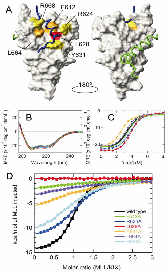
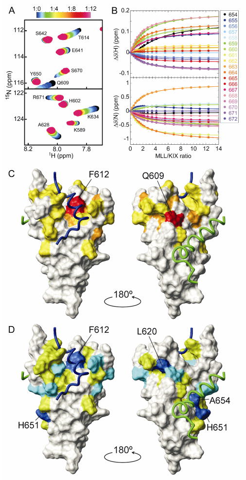
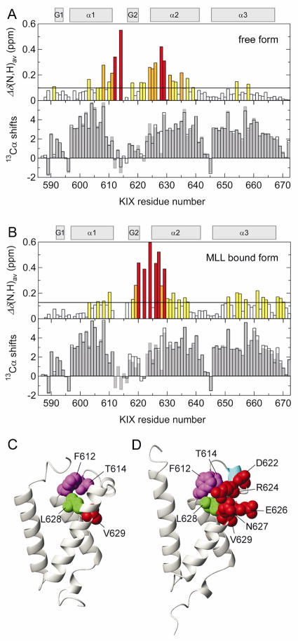
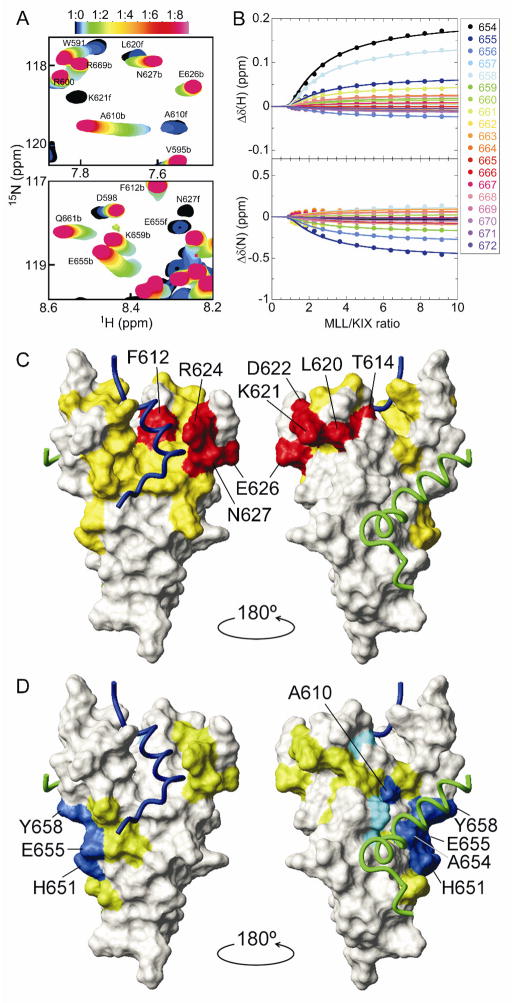
Physical interaction between the transactivation domain (TAD) of the mixed-lineage leukemia protein (MLL) and the KIX domain of the cyclic-AMP response element binding protein (CREB) binding protein (CBP) is necessary for MLL-mediated transcriptional activation. We show by alanine-scanning mutagenesis that hydrophobic surface residues of KIX, especially L628, are energetically important for binding the MLL TAD. NMR studies of the KIX-L628A mutant suggest that L628 plays a crucial role in conformational transitions at the MLL binding site, necessary for high affinity interactions with MLL. Unexpectedly, MLL also binds to the c-Myb/phosphorylated kinase-inducible domain of CREB (pKID) site of KIX, highlighting the complex nature of interactions involving intrinsically disordered transcriptional activators.


Figures

Figure 1
Figure 1
(A) Representation of the KIX structure in the ternary KIX·MLL·c-Myb complex (PDB 2AGH [6]). The six mutation sites are mapped onto the surface of KIX, color coded to indicate the effect of Ala substitution on Kd for MLL binding: yellow, 2-15× increase in Kd; orange, 25-40× increase; red, 100× increase. Backbones of the bound MLL (2842-2860 only) and c-Myb are shown in blue and green, respectively. The figure was prepared using MOLMOL [19]. (B) Far-UV CD spectra, (C) urea-induced equilibrium unfolding transition, and (D) MLL binding isotherms of the wild-type and mutant KIX. Color codes are shown in (D). In (C) and (D), continuous lines are the curves fitted to a two-state transition and a one-site binding model, respectively.
Figure 2
Figure 2
(A) Regions of the 1H-15N HSQC spectrum of the KIX-L628A mutant showing chemical shift changes upon titration with MLL. The cross-peak color changes gradually from black (free) to magenta according to the concentration ratio as shown in the bar above the figure. (B) 1H and 15N chemical shift titration curves for a subset of KIX-L628A resonances (colored points corresponding to residues according to the legend) upon titration with increasing amounts of MLL. The lines represent a global fit to the titration data, using a two-site binding model with Kd1 = 220 μM and Kd2 = 1.63 mM. (C,D) MLL binding sites on KIX-L628A. The weighted average chemical shift differences [ Δδ(N,H)av=(ΔδH)2+(ΔδN/5)2] for KIX-L628A amide resonances between the free form and the bound form, in which the primary (C) or secondary binding site (D) is occupied, are mapped onto the surface of KIX in the ternary complex, using colors to indicate changes in chemical shift greater than 2 × standard deviation (SD) from the mean (red and blue), between 1 and 2 × SD from the mean (orange and cyan), and between mean and 1 × SD from the mean (yellow and green).
Figure 3
Figure 3
(A, upper) Histogram showing Δδ(N,H)av between the wild-type and the L628A mutant of KIX in the free form. Color codes are the same as in Fig. 2. (A, lower) Histogram showing the secondary 13Cα chemical shifts calculated by subtraction of random coil values [20] from the experimental data for the wild-type KIX (open bars) and the L628A mutant (gray bars) in the free form. Regions showing positive secondary Cα chemical shifts indicate a helical conformation. Secondary structure elements in the ternary KIX·MLL·c-Myb complex [6] are depicted at the top. (B) The same as in (A), but in the presence of MLL (1:1 and 1:2 KIX:MLL ratios for the wild-type KIX and the L628A mutant, where 94% and 74% of KIX are in the MLL-bound form, respectively). (C,D) The KIX structures in the binary KIX·c-Myb complex (PDB 1SB0 [21]) and in the ternary KIX·MLL·c-Myb complex, respectively. Residues having large Δδ(N,H)av are shown by spheres of radius corresponding to the van der Waals radius of each atom. In (D), the G2 helix is colored cyan.
Figure 4
Figure 4
(A) Regions of the 1H-15N HSQC spectrum of wild-type KIX showing chemical shift changes upon titration with MLL. b and f indicate the peaks for the bound and free forms, respectively. (B) 1H and 15N chemical shift titration curves for a subset of KIX resonances upon titration with increasing amounts of MLL. The lines represent a global fit to the titration data, using a two-site binding model with Kd1 = 2.14 μM and Kd2 = 920 μM. (C,D) MLL binding sites on the wild-type KIX, as shown in Fig. 2C,D. Δδ(N,H)av for primary binding was obtained from the HSQC spectra at 1:0 and 1:1 KIX:MLL ratios. Δδ(N,H)av for secondary binding was obtained from a global fit of titration curves.

References

    1. Spiegelman BM, Heinrich R. Biological control through regulated transcriptional coactivators. Cell. 2004;119:157–167. - PubMed
    1. Goodman RH, Smolik S. CBP/p300 in cell growth, transformation, and development. Genes Dev. 2000;14:1553–1577. - PubMed
    1. Dyson HJ, Wright PE. Intrinsically unstructured proteins and their functions. Nat Rev Mol Cell Biol. 2005;6:197–208. - PubMed
    1. Radhakrishnan I, Perez-Alvarado GC, Parker D, Dyson HJ, Montminy MR, Wright PE. Solution structure of the KIX domain of CBP bound to the transactivation domain of CREB: a model for activator:coactivator interactions. Cell. 1997;91:741–752. - PubMed
    1. Goto NK, Zor T, Martinez-Yamout M, Dyson HJ, Wright PE. Cooperativity in transcription factor binding to the coactivator CREB-binding protein (CBP). The mixed lineage leukemia protein (MLL) activation domain binds to an allosteric site on the KIX domain. J Biol Chem. 2002;277:43168–43174. - PubMed

Publication types