. 2011 Jan 7;405(1):105-12.
doi: 10.1016/j.jmb.2010.10.010. Epub 2010 Oct 21.

Robust mechanosensing and tension generation by myosin VI

Affiliations

Robust mechanosensing and tension generation by myosin VI

Peiying Chuan et al. J Mol Biol. .

Abstract

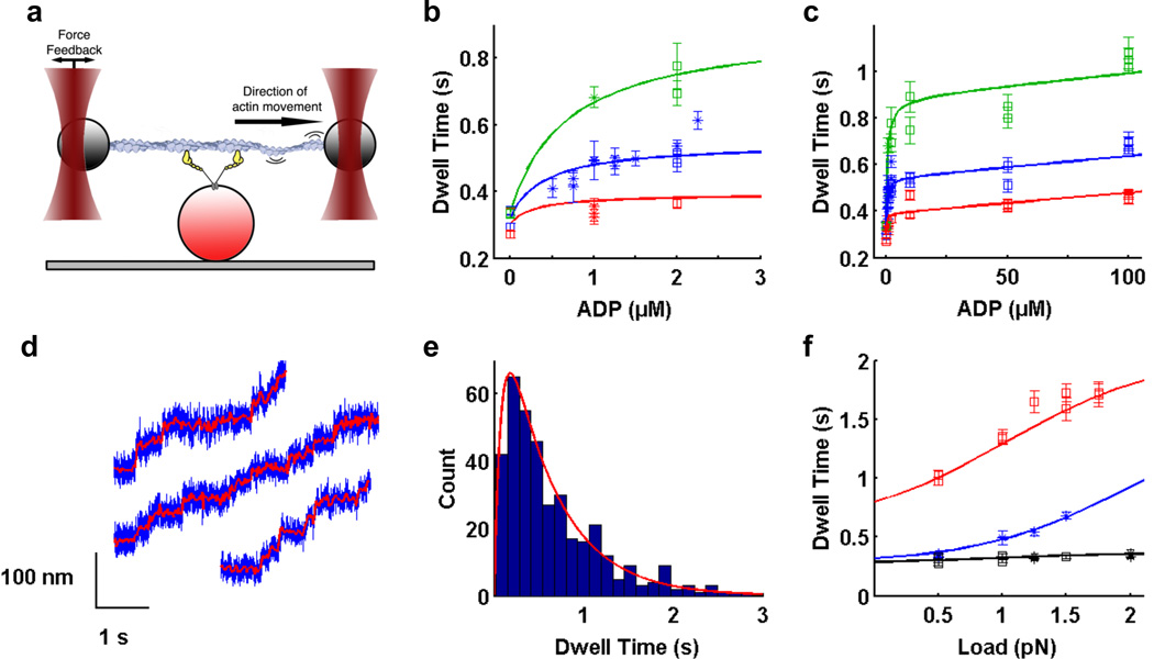
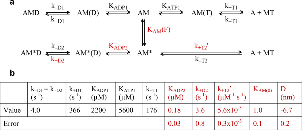
Myosin VI is a molecular motor that is thought to function both as a transporter and as a cytoskeletal anchor in vivo. Here we use optical tweezers to examine force generation by single molecules of myosin VI under physiological nucleotide concentrations. We find that myosin VI is an efficient transporter at loads of up to ∼2 pN but acts as a cytoskeletal anchor at higher loads. Our data and the resulting model are consistent with an indirect coupling of global structural motions to nucleotide binding and release. The model provides a mechanism by which load may regulate the dual functions of myosin VI in vivo. Our results suggest that myosin VI kinetics are tuned such that the motor maintains a consistent level of mechanical tension within the cell, a property potentially shared by other mechanosensitive proteins.


Figures

Figure 1
Figure 1. Optical trapping data and regression curves from kinetic model
a) Optical trapping assay. Two polystyrene beads attached to ends of an actin filament are held in independently controlled optical traps. The actin filament is brought into contact with a surface platform bead-bound myosin VI molecule. Feedback control on one trap maintains the force applied on the myosin molecule as the motor walks along actin. b,c) Dependence of dwell time on b) low ADP concentrations up to 2.5 µM; c) ADP concentrations up to 100 µM, in the presence of 2 mM ATP (for zero ADP only) or 1.5 mM ATP and 0.5 pN (red), 1.0 pN (blue) and 1.5 pN (green) load. Within each condition, each data point represents a different myosin VI molecule. Squares are our measured dwell times, asterisks are data from ref. . Errors for dwell times were obtained by bootstrapping with 1000 replicates to get the standard error of the mean (SEM). d) Stepping traces of myosin VI in the optical trap in the presence of 1.5 mM ATP, 100 µM ADP and 1 pN load. Filtered bead positions (red traces) are overlaid on raw bead positions (blue traces). e) Dwell time histogram for one molecule under the same conditions as in d). Average dwell time is 0.69 ± 0.03 s. Red curve is the predicted dwell time distribution based on our model. f) Dependence of dwell time on load in the presence of 2 mM ATP (black), 1.5 mM ATP plus 1 µM ADP (blue), and 0.1 mM ATP (red). b,c,f) Best fit curves to dwell time data from global weighted least squares regression (solid lines). Refer to Supplemental Information for details of data analysis.
Figure 2
Figure 2. Kinetic model and parameters for myosin VI under load
a) Kinetic scheme for myosin VI under load. Myosin exists in two states, M and M*. AM represents the myosin VI rear head (M) bound to actin (A), and D and T represent ADP and ATP respectively. KAM(F) = KAM(0) exp(−FD/kBT) is the force-sensitive equilibrium constant between AM and AM*. All other uppercase K’s are affinity constants, and lowercase k’s are rate constants. b) Previously determined parameters, that were fixed in the regression are in black. Values from the regression and their errors are in red. Each data point was weighted by the inverse of its squared standard error of the mean. Errors on the parameters were determined by assuming that each data point follows a normal distribution centered at the mean with a standard deviation of SEM, and performing a parametric bootstrap with 2000 replicates. Refer to Supplemental Information for details of regression analysis.
Figure 3
Figure 3. The dual functions of myosin VI are regulated by load in vivo
a) Dependence of dwell time on load under physiological 1.5 mM ATP and 100 µM ADP (data used in regression: blue points; model: blue curve). The model correctly predicts dwell times at higher forces (red). Within each condition, each data point represents a different myosin VI molecule. Cartoons depict myosin VI motors stepping along actin while attached via adapter proteins (grey ovals) to a membrane. At low loads, the rate of ADP binding (red arrow) is low (leftmost cartoon). The rear head predominantly binds ATP (green arrow), resulting in 2–3 steps s−1. At moderate forces ADP competes with ATP for binding to the rear head, slowing the motor to less than 1 step s−1 (center). At forces greater than ~2.5 pN, the stepping rate decreases rapidly as ADP out-competes ATP for binding (right). The motor takes slow backsteps before detaching from actin (see Supplemental Information). In this way, M6 is able to maintain tension at ~2 pN and relieve forces greater than ~2.5 pN.

References

    1. Lin HP, Chen HM, Wei SY, Chen LY, Chang LH, Sun YJ, et al. Cell adhesion molecule Echinoid associates with unconventional myosin VI/Jaguar motor to regulate cell morphology during dorsal closure in Drosophila. Dev Biol. 2007;311:423–433. - PubMed
    1. Mohiddin SA, Ahmed ZM, Griffith AJ, Tripodi D, Friedman TB, Fananapazir L, et al. Novel association of hypertrophic cardiomyopathy, sensorineural deafness, and a mutation in unconventional myosin VI (MYO6) J Med Genet. 2004;41:309–314. - PMC - PubMed
    1. Hertzano R, Shalit E, Rzadzinska AK, Dror AA, Song L, Ron U, et al. A Myo6 mutation destroys coordination between the myosin heads, revealing new functions of myosin VI in the stereocilia of mammalian inner ear hair cells. PLoS Genet. 2008;4 e1000207. - PMC - PubMed
    1. Buss F, Kendrick-Jones J. How are the cellular functions of myosin VI regulated within the cell? Biochem Biophys Res Commun. 2008;369:165–175. - PMC - PubMed
    1. Cai Y, Sheetz MP. Force propagation across cells: mechanical coherence of dynamic cytoskeletons. Curr Opin Cell Biol. 2009;21:47–50. - PMC - PubMed

Publication types

LinkOut - more resources