. 2011 Jan 1;50(1):55-65.
doi: 10.1016/j.freeradbiomed.2010.10.689. Epub 2010 Oct 21.

Marked changes in endogenous antioxidant expression precede vitamin A-, C-, and E-protectable, radiation-induced reductions in small intestinal nutrient transport

Affiliations

Marked changes in endogenous antioxidant expression precede vitamin A-, C-, and E-protectable, radiation-induced reductions in small intestinal nutrient transport

Marjolaine Roche et al. Free Radic Biol Med. .

Abstract

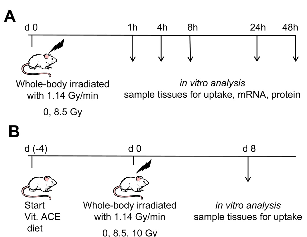
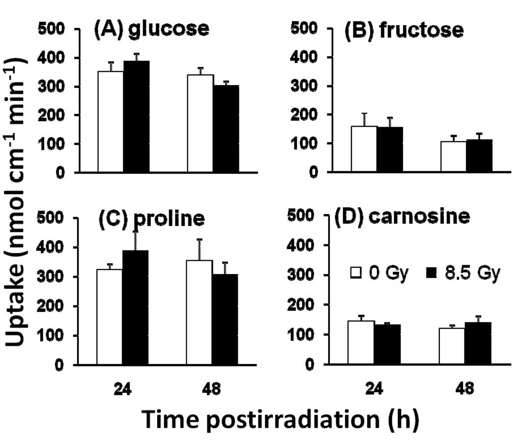
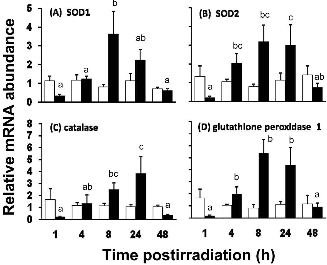
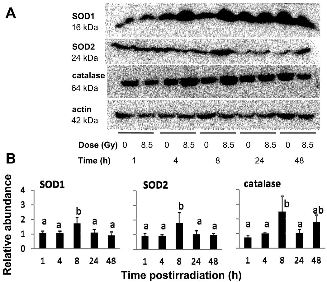
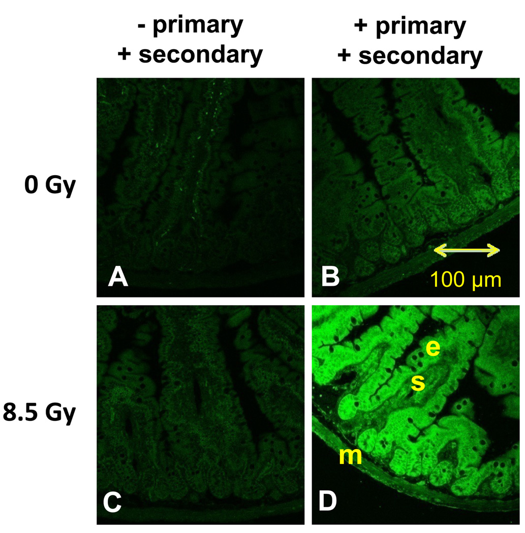
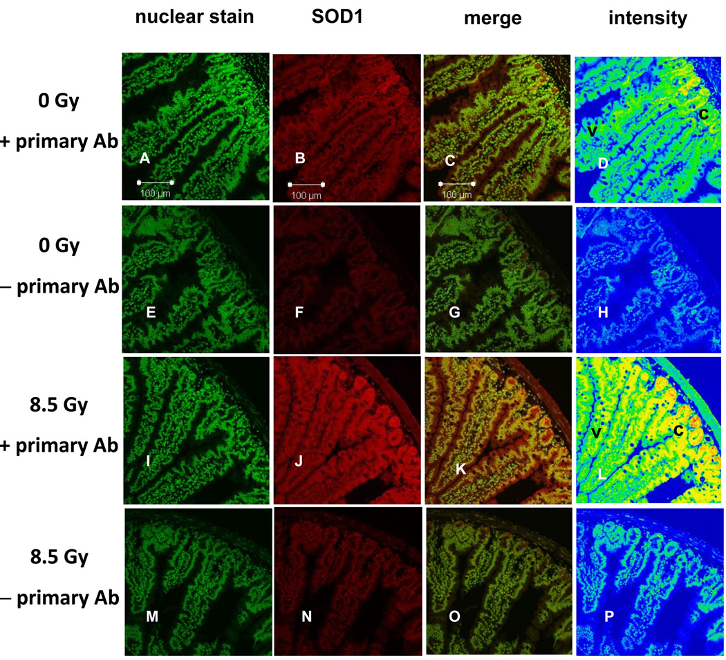
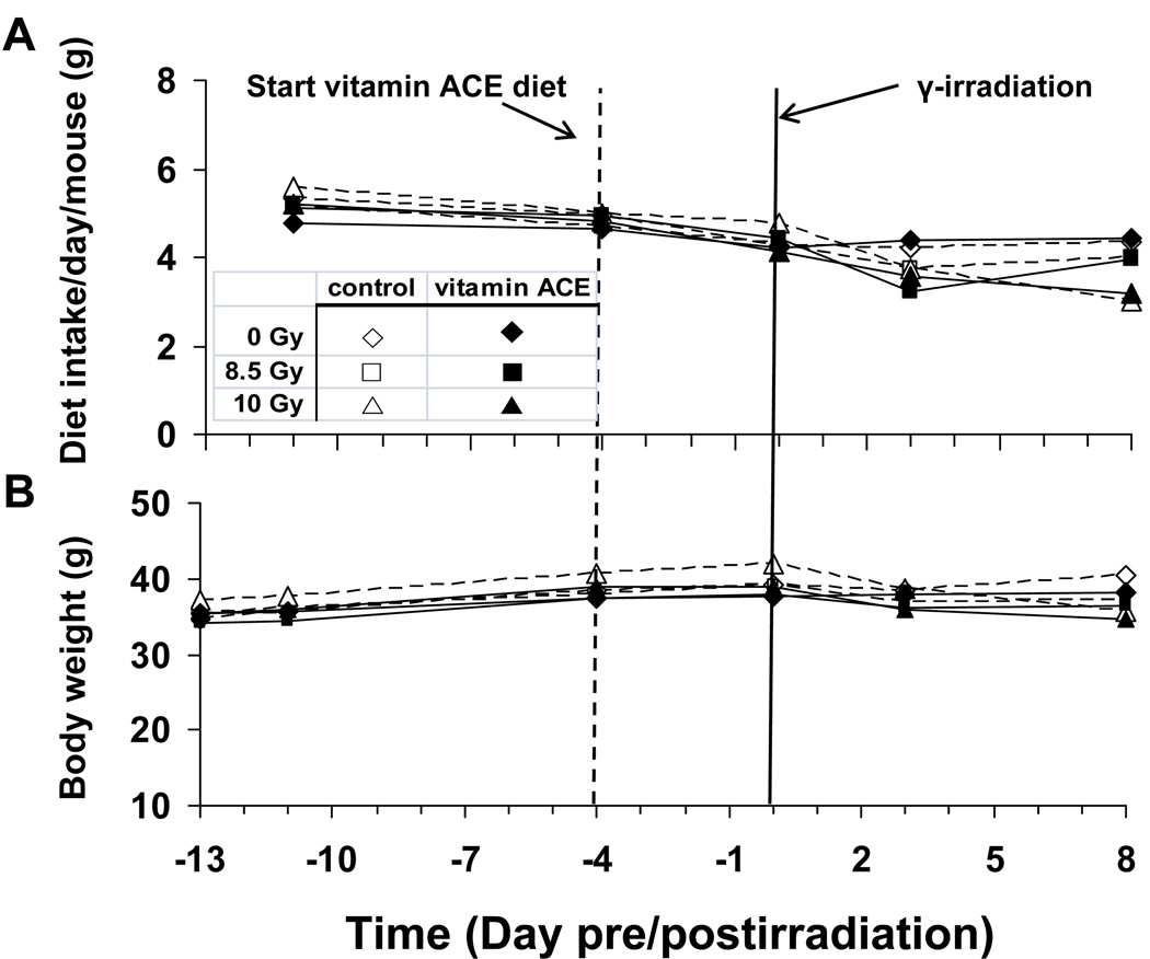
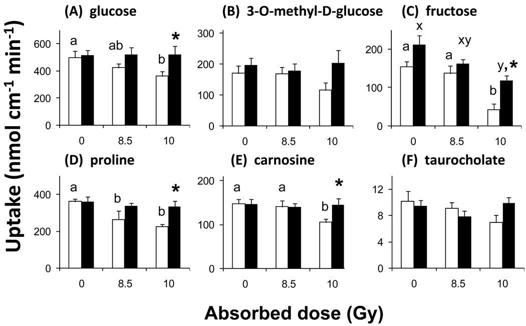
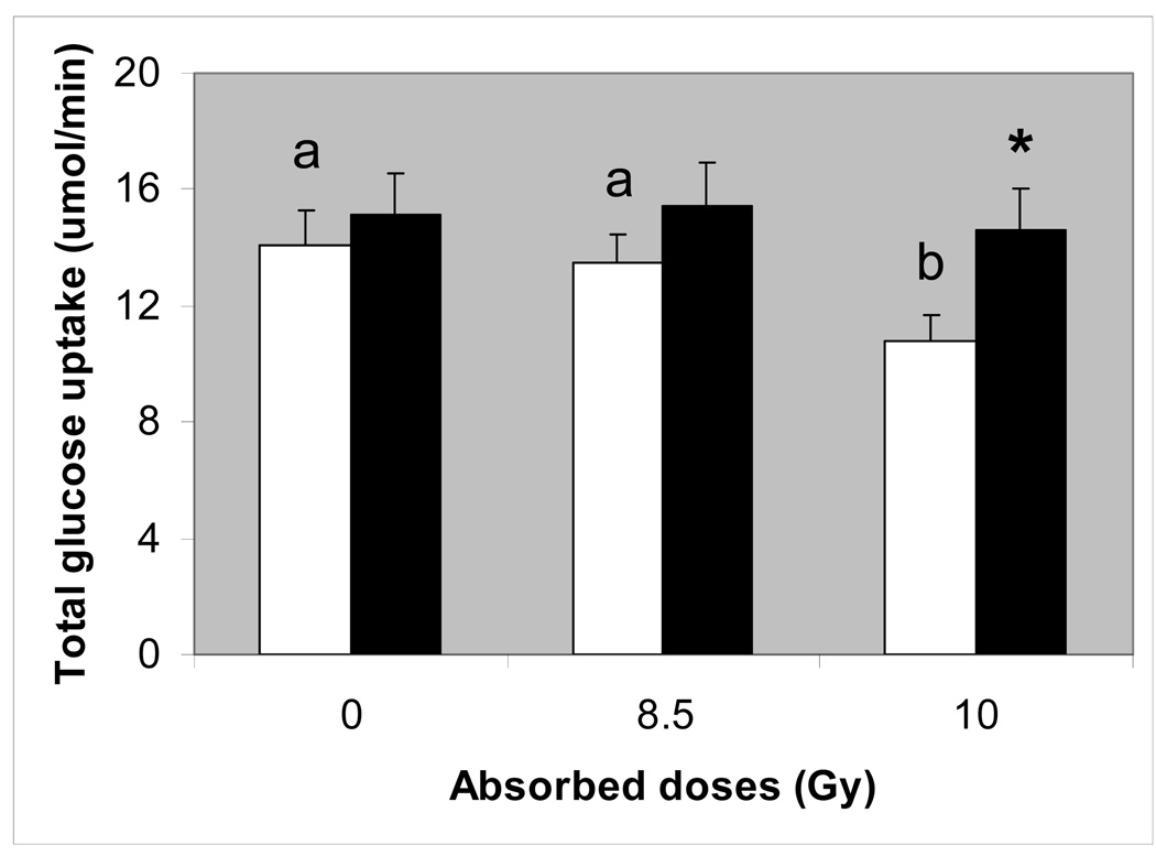
Rapidly proliferating epithelial crypt cells of the small intestine are susceptible to radiation-induced oxidative stress, yet there is a dearth of data linking this stress to expression of antioxidant enzymes and to alterations in intestinal nutrient absorption. We previously showed that 5-14 days after acute γ-irradiation, intestinal sugar absorption decreased without change in antioxidant enzyme expression. In the present study, we measured antioxidant mRNA and protein expression in mouse intestines taken at early times postirradiation. Observed changes in antioxidant expression are characterized by a rapid decrease within 1h postirradiation, followed by dramatic upregulation within 4h and then downregulation a few days later. The cell type and location expressing the greatest changes in levels of the oxidative stress marker 4HNE and of antioxidant enzymes are, respectively, epithelial cells responsible for nutrient absorption and the crypt region comprising mainly undifferentiated cells. Consumption of a cocktail of antioxidant vitamins A, C, and E, before irradiation, prevents reductions in transport of intestinal sugars, amino acids, bile acids, and peptides. Ingestion of antioxidants may blunt radiation-induced decreases in nutrient transport, perhaps by reducing acute oxidative stress in crypt cells, thereby allowing the small intestine to retain its absorptive function when those cells migrate to the villus days after the insult.


Figures

Fig. 1
Fig. 1
Design of experiments wherein mice were acutely irradiated with low-LET γ-rays and sacrificed at different times postirradiation. (A) Mice were fed control diet, irradiated, and sacrificed 1 – 48 h postirradiation. (B) Mice were fed control diet or diet supplemented with vitamin A, C, and E. Arrows refer to times postirradiation when mice were sacrificed. Day 8 was chosen for B based on a previous experiment tracking the time course of radiation-induced reductions in nutrient transport.
Fig. 2
Fig. 2
In vitro measurements of nutrient uptake rate in isolated jejunum of unirradiated (open bars) and irradiated (8.5 Gy, filled bars) mice 24 and 48 h postirradiation. (A) D-Glucose, (B) D-fructose, (C) proline, and (D) carnosine uptakes per cm. Results are means ± SE (n = 6). By two way ANOVA, there were no significant effects of radiation and of time postirradiation on all nutrient uptakes (see text for P values).
Fig. 3
Fig. 3
Relative mRNA abundance of antioxidant enzymes in isolated jejunum of unirradiated (open bars) and irradiated (8.5 Gy, filled bars) mice. (A) SOD1, (B) SOD2, (C) catalase, and (D) glutathione peroxidase 1 as measured by real time PCR. mRNA was collected from the mucosa of the proximal small intestine of mice fed with control diet and sacrificed 1, 4, 8, 24 and 48 h post-irradiation. Results are means ± SE (n = 6). Filled bars with different superscripts are significantly different (P < 0.05 by one-way ANOVA). Thus, there is a marked decrease in levels of mRNA at 1irradiation, followed by a dramatic increase, and then a decline to levels similar to unirradiated mice. Open bars are similar throughout, indicating that expression in unirradiated mice did not change over time.
Fig. 4
Fig. 4
(A) Effect of γ-irradiation on protein abundance of the antioxidant enzymes SOD1, SOD2, and catalase as determined by western blotting of mucosal homogenates. Homogenates were obtained from the jejunum of mice fed with control diet and sacrificed 1, 4, 8, 24 and 48 h postirradiation. (B) Mean relative antioxidant enzyme protein levels based on densitometry scans of western blots. Results are the ratio of the densities corresponding to 8.5Gy and 0Gy from homogenates obtained from the same trial. Results are means ± SE (n = 5 for SOD1 n = 5 for SOD2, and n = 4 for catalase). Bars with different superscripts are significantly different (P < 0.05 by one-way ANOVA).
Fig. 5
Fig. 5
Immunolocalization of the oxidative stress biomarker 4HNE in the small intestine of mice 8 h after acute whole-body irradiation with 8.5 Gy. (A) Intestinal section from unirradiated mice probed with a secondary (Alexa 488 goat antirabbit) antibody but no primary (rabbit anti-4HNE) antibody. (B) Section from unirradiated mice probed with both primary and secondary antibodies. (C) Section from irradiated mice probed with a secondary antibody only. (D) High levels of 4HNE were found in the intestine of irradiated mice probed with primary and secondary antibodies. Epithelial cells (e) lining the mucosa seemed to have greater amounts of 4HNE than the submucosal (s) or muscle (m) layers.
Fig. 6
Fig. 6
Immunolocalization of SOD1 (red) in the small intestine after acute whole-body irradiation with 0 Gy and 8.5 Gy. Sections in column 1 were stained with Sytox Green (nuclear stain). Column 3 represents merged images from columns 1 and 2, while column 4 depicts staining intensity where blue = low, green = modest, yellow = high and red = highest intensity. Sections from unirradiated mice with primary antibody (row 1, panels A – D) and without (row 2, panels E – H). Irradiated mice with primary antibody (row 3, panels I – L) and without (row 4, panels M – P). SOD1 expression is greater in irradiated mice (compare panels B, C with J, K), confirming western blots in Fig. 4. Control panels H and P show no difference in intensity of staining of cytosol and nuclei along the crypt-villus axis when no anti-SOD1 is present. Before irradiation, endogenous SOD1 expression seems already greater in the crypt (top right, c) as opposed to upper villus (v) regions (panel D). After irradiation, there is a dramatic increase in SOD1 throughout the mucosa, and little increase in SOD1 in the submucosal and muscle layers (panel L). SOD1 expression seems greatest in the crypt region (c, panel, L) of intestines from irradiated mice.
Fig. 7
Fig. 7
As diagrammed in Fig. 1B, this is the effect of diet and radiation dose on (A) dietary intake (g per day per mouse), and (B) body weight as a function of time pre and postirradiation. Here, the mice were irradiated with 0 (control), 8.5 or 10 Gy at day 0 (see solid vertical line). The mice were housed one per cage and fed with either control (open symbols) or vitamin ACE enriched diet (closed symbols). Mean results are plotted, n = 6. Standards errors have been omitted for clarity. Mice were fed with the control diet from day −13 to day −4. Then, half of the mice were switched to the vitamin ACE-supplemented diet (dashed vertical line). Diet and radiation dose had modest effects on feeding rate and body weight.
Fig.8
Fig.8
In vitro measurements of (A) D-Glucose, (B) 3-O-methyl-D-glucose, (C) D-fructose, (D) proline, (E) carnosine uptake rate in the jejunum of mice irradiated with 0, 8.5, or 10 Gy and sacrificed 8 d postirradiation. Taurocholate (F) uptake was determined in the distal ileum. Mice were fed AIN-76A diet (control, open bars) or an AIN-76A diet supplemented with a cocktail of vitamins A, C, and E (filled bars). Results are means (± SE) of 6 independent experiments. Open bars with different letter superscripts are significantly different from other open bars (P < 0.05 by one-way ANOVA), suggesting that increasing radiation dose reduce uptake rate. With the exception of fructose, filled bars did not decrease with increasing radiation dose, suggesting that consumption of the vitamin ACE-supplemented diet prevented the radiation-induced reduction in uptake. Filled bars with asterisks are significantly greater (P < 0.05 by one-way ANOVA) than open bars at the same radiation dose, suggesting that uptake is greater in tissues from vitamin-treated mice. Similar results are obtained when uptakes are expressed per mg of intestine.
Fig. 9
Fig. 9
Total glucose uptake for the entire small intestine, estimated by integrating uptakes from the duodenal, jejuna and distal regions. Open bars with different letter superscripts are significantly different (P < 0.05 by one-way ANOVA), suggesting that increasing radiation dose reduced uptake rate. Filled bars did not change with radiation dose, suggesting that consumption of the vitamin ACE-supplemented diet prevented the radiation-induced reduction in uptake. The filled bar with asterisk is significantly greater (P < 0.05 by one-way ANOVA) than the open bar at 10 Gy, suggesting that uptake was greater in tissues from vitamin-treated mice.

References

    1. Potten CS, Merritt A, Hickman J, Hall P, Faranda A. Characterization of radiation-induced apoptosis in the small intestine and its biological implications. Int J Radiat Biol. 1994;65:71–78. - PubMed
    1. Somosy Z, Horvath G, Telbisz A, Rez G, Palfia Z. Morphological aspects of ionizing radiation response of small intestine. Micron. 2002;33:167–178. - PubMed
    1. Miholic J, Vogelsang H, Schlappack O, Kletter K, Szepesi T, Moeschl P. Small bowel function after surgery for chronic radiation enteritis. Digestion. 1989;42:30–38. - PubMed
    1. Hauer-Jensen M, Wang J, Boerma M, Fu Q, Denham JW. Radiation damage to the gastrointestinal tract: mechanisms, diagnosis, and management. Curr Opin Support Palliat Care. 2007;1:23–29. - PubMed
    1. Strup-Perrot C, Mathe D, Linard C, Violot D, Milliat F, Francois A, Bourhis J, Vozenin-Brotons MC. Global gene expression profiles reveal an increase in mRNA levels of collagens, MMPs, and TIMPs in late radiation enteritis. Am J Physiol Gastrointest Liver Physiol. 2004;287:G875–G885. - PubMed

Publication types

MeSH terms