. 2011 Jan;50(1):87-98.
doi: 10.1016/j.yjmcc.2010.10.010. Epub 2010 Oct 30.

PKA and Epac synergistically inhibit smooth muscle cell proliferation

Affiliations

PKA and Epac synergistically inhibit smooth muscle cell proliferation

Richard C Hewer et al. J Mol Cell Cardiol. 2011 Jan.

Abstract

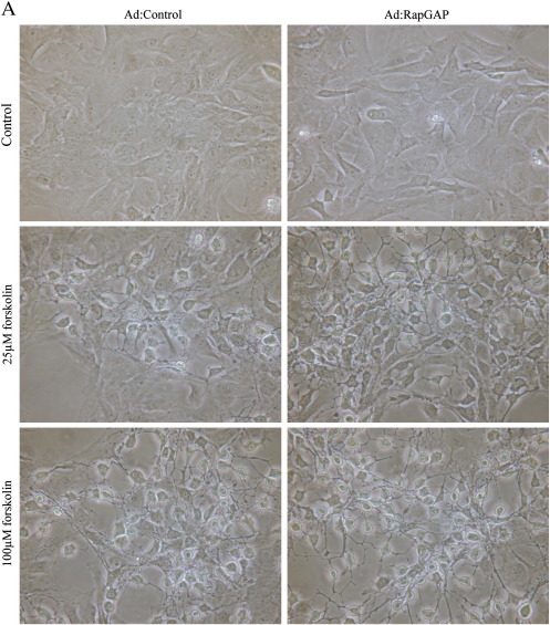
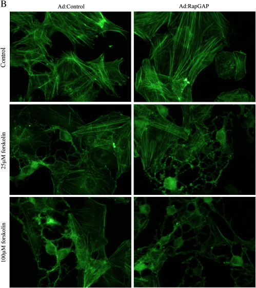
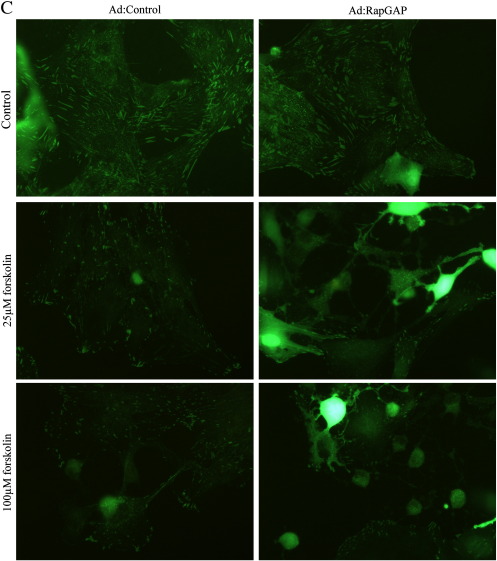
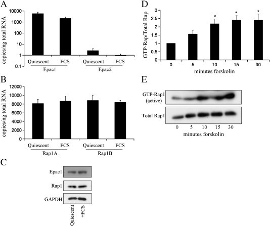
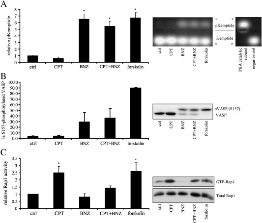
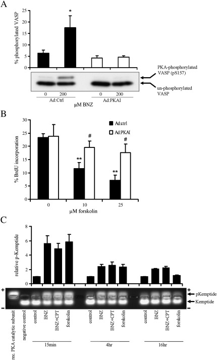
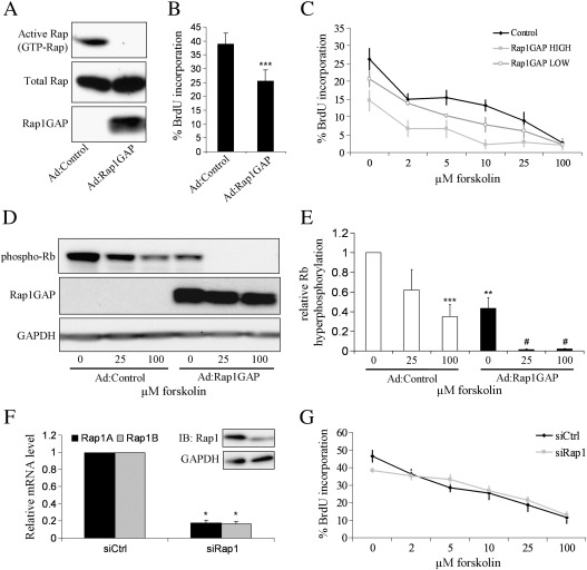
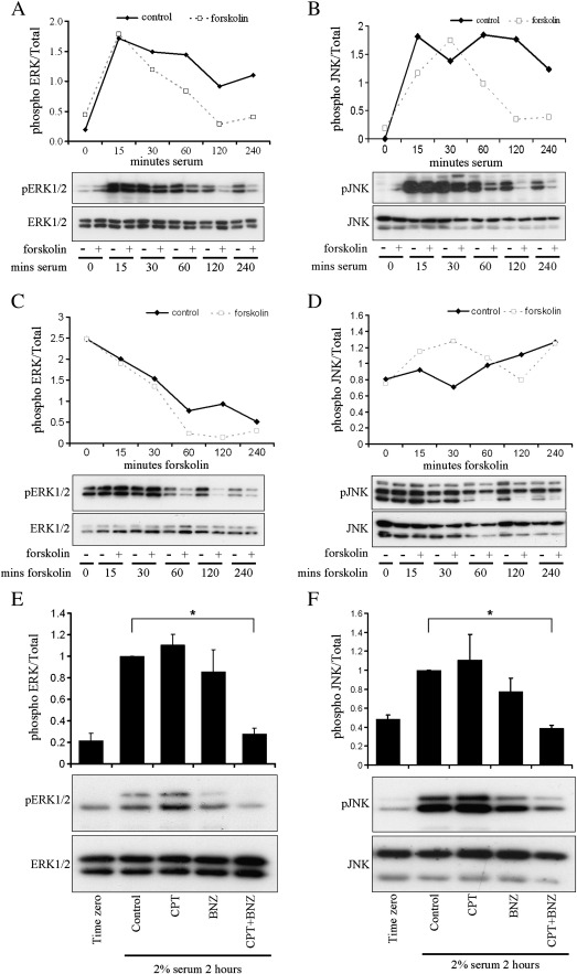
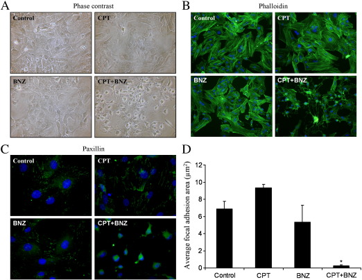
Cyclic AMP signalling promotes VSMC quiescence in healthy vessels and during vascular healing following injury. Cyclic AMP inhibits VSMC proliferation via mechanisms that are not fully understood. We investigated the role of PKA and Epac signalling on cAMP-induced inhibition of VSMC proliferation. cAMP-mediated growth arrest was PKA-dependent. However, selective PKA activation with 6-Benzoyl-cAMP did not inhibit VSMC proliferation, indicating a requirement for additional pathways. Epac activation using the selective cAMP analogue 8-CPT-2'-O-Me-cAMP, did not affect levels of hyperphosphorylated Retinoblastoma (Rb) protein, a marker of G1-S phase transition, or BrdU incorporation, despite activation of the Epac-effector Rap1. However, 6-Benzoyl-cAMP and 8-CPT-2'-O-Me-cAMP acted synergistically to inhibit Rb-hyperphosphorylation and BrdU incorporation, indicating that both pathways are required for growth inhibition. Consistent with this, constitutively active Epac increased Rap1 activity and synergised with 6-Benzoyl-cAMP to inhibit VSMC proliferation. PKA and Epac synergised to inhibit phosphorylation of ERK and JNK. Induction of stellate morphology, previously associated with cAMP-mediated growth arrest, was also dependent on activation of both PKA and Epac. Rap1 inhibition with Rap1GAP or siRNA silencing did not negate forskolin-induced inhibition of Rb-hyperphosphorylation, BrdU incorporation or stellate morphology. This data demonstrates for the first time that Epac synergises with PKA via a Rap1-independent mechanism to mediate cAMP-induced growth arrest in VSMC. This work highlights the role of Epac as a major player in cAMP-dependent growth arrest in VSMC.


Figures

Supplemental Fig. 1
Supplemental Fig. 1
Epac and PKA synergise to inhibit VSMC proliferation. A, Asynchronous VSMC were stimulated with 200 μM BNZ, with or without 200 μM CPTSp stimulation, for 24 h with the last 6 h in the presence of 10 μM BrdU and analysed for BrdU incorporation, n = 6. * indicates p < 0.05 versus control.
Supplemental Fig. 2
Supplemental Fig. 2
Inhibition of Rap does not negate the effects of cAMP on morphology. Asynchronous VSMC were infected with an adenovirus encoding Rap1GAP and cells were stimulated with 25 and 100 μM forskolin. A, Phase contrast images showing cell morphology. B, Phalloidin staining for F-actin. C, Paxillin staining representing focal adhesions.
Supplemental Fig. 2
Supplemental Fig. 2
Inhibition of Rap does not negate the effects of cAMP on morphology. Asynchronous VSMC were infected with an adenovirus encoding Rap1GAP and cells were stimulated with 25 and 100 μM forskolin. A, Phase contrast images showing cell morphology. B, Phalloidin staining for F-actin. C, Paxillin staining representing focal adhesions.
Fig. 1
Fig. 1
The Epac–Rap pathway is expressed and functional in rat VSMC. VSMC were rendered quiescent by serum deprivation and stimulated with FCS as indicated: A and B, Epac1, Epac2, Rap1A and Rap1B mRNA copy number was calculated by quantitative RT-PCR, n = 5; C, Epac1 and Rap1 protein expression was verified by western blotting. D, VSMC were stimulated with 100 μM forskolin for indicated times, and assayed for Rap1 activity by Ral-GDS pulldown, n = 6. E, A representative western blot is shown. * indicates p < 0.01 versus 0 min.
Fig. 2
Fig. 2
Selective activation of PKA and Epac signalling with N6-Benzoyl-cAMP and 8-CPT-2′-O-Me-cAMP, respectively. VSMC were serum deprived for 24 h and stimulated with 200 μM BNZ, CPT, BNZ plus CPT or 100 μM forskolin for 30 min as indicated. A, Cell lysates were analysed for PKA activity by kemptide phosphorylation assay. Upper bands represent PKA phosphorylated kemptide while lower bands are un-phosphorylated kemptide. Recombinant PKA catalytic subunit and lysis buffer were included in the assay as positive and negative controls respectively. B, Cell lysates were analysed for phospho-VASP (slower migrating band) by western blotting with a total VASP antibody, n = 3. C, Rap1 activity was quantified by Ral-GDS pulldown, n = 6. * indicates p < 0.05 versus control.
Fig. 3
Fig. 3
Epac and PKA synergise to inhibit VSMC cell-cycle progression. Asynchronous VSMC were stimulated with 200 μM BNZ, 200 μM CPT, 200 μM BNZ plus 200 μM CPT, or 200 μM db-cAMP: A, for 24 h with the last 6 h in the presence of 10 μM BrdU and analysed for BrdU incorporation, n = 6; B and C, for 18 h and cell lysates analysed by western blotting as indicated, n = 3. * indicates p < 0.05, ** indicates p < 0.01, *** indicates p < 0.001 versus control.
Fig. 4
Fig. 4
PKA is required but not sufficient to inhibit VSMC cell-cycle progression. VSMC were infected with either Ad:Control or Ad:PKAI at 2 × 108 pfu/ml. A, 40 h later, cells were rendered quiescent by serum deprivation for a further 6 h followed by stimulation with 200 μM BNZ for 15 min. Total cell lysates were analysed for non-phosphorylated (lower band) and phosphorylated (upper band) VASP by western blotting, n = 3. B, Asynchronous VSMC were stimulated with forskolin for 24 h with the last 6 h in the presence of 10 μM BrdU. BrdU incorporation was quantified by immuno-staining for BrdU and counting of positively stained nuclei, n = 7. C, Asynchronous VSMC were stimulated for the indicated times with 200 μM BNZ, 200uM CPT, 200 μM BNZ plus 200 μM CPT, or 100 μM forskolin, and analysed for PKA activity by kemptide phosphorylation assay. Upper bands represent PKA phosphorylated kemptide while lower bands are un-phosphorylated kemptide. Recombinant PKA catalytic subunit and lysis buffer were included in the assay as positive and negative controls respectively. * indicates p < 0.05, ** indicates p < 0.01 versus Ad:Control alone. # indicates p < 0.05 versus corresponding dose in Ad:Control.
Fig. 5
Fig. 5
Constitutively active Epac synergises with PKA to inhibit VSMC cell-cycle progression. Asynchronous VSMC were infected with an adenovirus encoding ΔcAMP-Epac as indicated. A, Rap1 activity in expressing and non-expressing cells was detected by Ral-GDS pulldown, n = 4. Cells were stimulated with 200 μM BNZ: B, for 24 h with the last 6 h in the presence of 10 μM BrdU and analysed for BrdU incorporation, n = 4; C, for 18 h and cell lysates were analysed by western blotting for Rb-hyperphosphorylation, n = 6. ** indicates p < 0.01 versus Ad:ΔcAMP-Epac.
Fig. 6
Fig. 6
Inhibition of Rap1 does not negate the effects of cAMP on cell-cycle progression. Where indicated, asynchronous VSMC were infected with an adenovirus encoding Rap1GAP at 108 pfu/ml (high) or 2 × 107 pfu/ml (low) (A–E), or transfected with siRNA specific for Rap1 as described (F, G). High dose of Ad:Rap1GAP was used unless otherwise indicated. VSMC were stimulated with the indicated forskolin doses for 18 h. A, Rap1GAP activity was confirmed by Rap1 activity assay. B, VSMC were incubated for 6 h in the presence of 10 μM BrdU and analysed for BrdU incorporation, n = 9. C, VSMC were stimulated with the forskolin doses indicated for a further 6 h in the presence of 10 μM BrdU and analysed for BrdU incorporation, n = 3; D and E, cell lysates were analysed by western blotting as indicated, n = 5. F, 24 h post transfection, mRNA levels of Rap1A and Rap1B were quantified by RT-PCR, n = 4; F (inset), and cell lysates were analysed by western blotting with a pan-Rap1A/B antibody; G, VSMC were stimulated with the indicated forskolin doses a further 6 h in the presence of 10 μM BrdU and analysed for BrdU incorporation, n = 6. * indicates p < 0.05 versus siCtrl. ** indicates p < 0.01, *** indicates p < 0.001 versus Ad:Control. # indicates p < 0.05, ## indicates p < 0.01 versus Ad:Rap1GAP.
Fig. 7
Fig. 7
Epac and PKA synergise to inhibit phosphorylation of ERK1/2 and JNK. A and B, Effect of forskolin on acute serum stimulated ERK1/2 and JNK phosphorylation. VSMC were rendered quiescent by serum deprivation for 24 h and pre-treated with 25 μM forskolin as indicated for 30 min before stimulation with 10% serum in the presence of 25 μM forskolin for the timepoints (minutes) indicated. C and D, Effect of forskolin on ERK1/2 and JNK phosphorylation in cells grown continuously in the presence of serum. VSMC were stimulated with 25 μM forskolin in the presence of 10% serum for the timepoints (minutes) indicated. E and F, Effect of PKA and Epac-selective analogues on acute serum stimulated ERK1/2 and JNK phosphorylation. VSMC were rendered quiescent for 24 h and pre-treated with 200 μM of the indicated cAMP analogues before 2% serum stimulation for the indicated timepoints (minutes). Levels of phosphorylated and total ERK1/2 and JNK were quantified by western blotting. * indicates p < 0.05; ANOVA with Student–Newman–Keuls post test on logged data.
Fig. 8
Fig. 8
Epac and PKA synergise to induce stellate morphology. Asynchronous VSMC were stimulated with 200 μM BNZ, CPT or BNZ plus CPT as indicated. A, Phase contrast images showing cell morphology. B, Phalloidin staining for F-actin. C, Paxillin staining representing focal adhesions, D, the area of which was quantified using NIH ImageJ, n = 3. * indicates p < 0.05 versus control.

References

    1. Izzard T., Taylor C., Birkett S., Jackson C., Newby A. Mechanisms underlying maintenance of smooth muscle cell quiescence in rat aorta: role of the cyclin dependent kinases and their inhibitors. Cardiovasc Res. 2002;52:242–252. - PubMed
    1. Lewis C.D., Olson N.E., Raines E.W., Reidy M.A., Jackson C.L. Modulation of smooth muscle proliferation in rat carotid artery by platelet-derived mediators and fibroblast growth factor-2. Platelets. 2001;12:352–353. - PubMed
    1. Hedin U., Roy J., Tran P.K. Control of smooth muscle cell proliferation in vascular disease. Curr Opin Cell Biol. 2004;15:559–565. - PubMed
    1. Wu Y., Bond M., Sala-Newby G., Newby A. Altered S-phase kinase-associated protein-2 levels are a major mediator of cyclic nucleotide-induced inhibition of vascular smooth muscle cell proliferation. Circ Res. 2006;98:1141–1150. - PubMed
    1. Indolfi C., Avvedimento E.V., DiLorenzo E., Esposito G., Rapacciuolo A., Giuliano P. Activation of cAMP-PKA signaling in vivo inhibits smooth muscle cell proliferation induced by vascular injury. Nat Med. 1997;3:775–779. - PubMed

Publication types

MeSH terms