

Very small embryonic-like stem cells in cardiovascular repair
- PMID: 20971132
- PMCID: PMC3005032
- DOI: 10.1016/j.pharmthera.2010.09.012
Very small embryonic-like stem cells in cardiovascular repair
Abstract
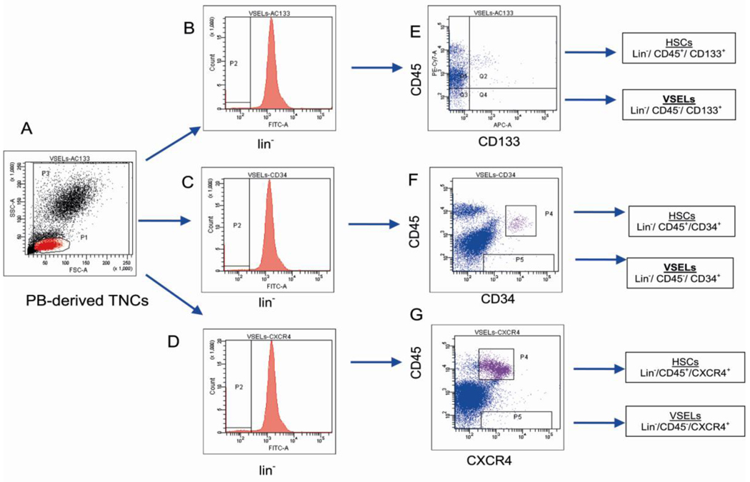
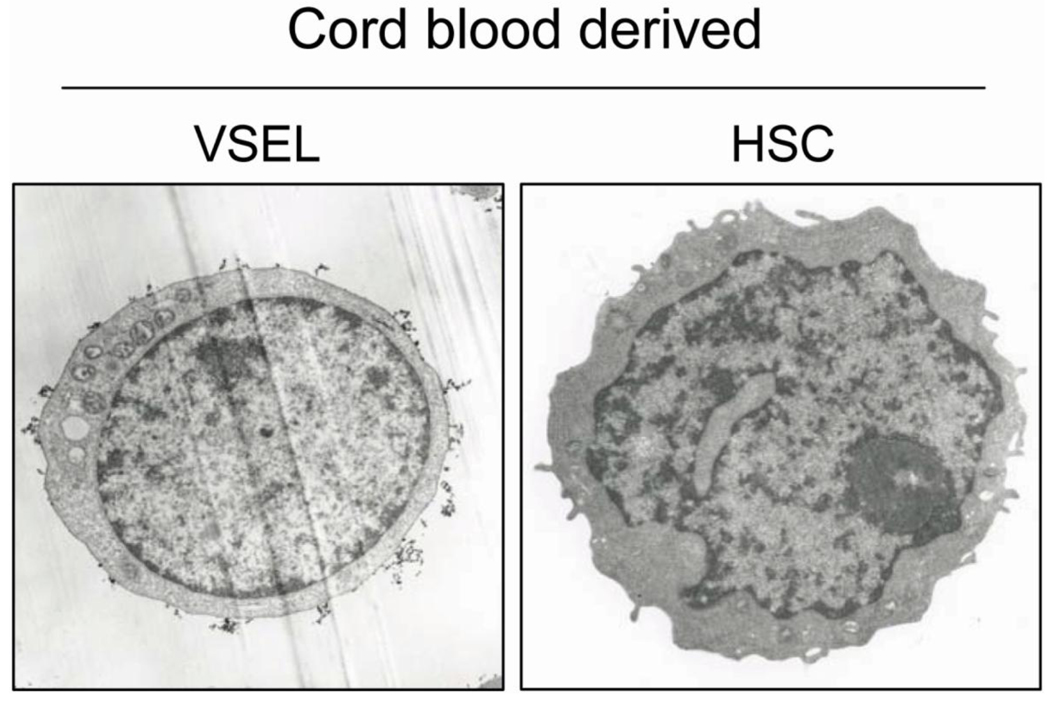
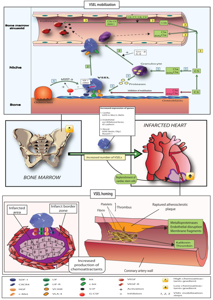
Adult bone marrow (BM) harbors several small populations of cells which may contribute to cardiac and endothelial repair, such as endothelial progenitor cells (EPCs), mesenchymal stromal cells (MSCs) and very small embryonic-like cells (VSELs) expressing several markers of pluripotent stem cells (PSCs), such as Oct-4, Nanog and SSEA-1. Such cells were identified in mice bone marrow, peripheral blood and solid organs as well as in umbilical cord blood (UCB) and peripheral blood (PB) in humans. The adult BM-derived VSELs may undergo differentiation into cells derived for all three germ layers, including cardiomyocytes and vascular endothelial cells. VSELs can be isolated using a multiparameter live cell sorting technique with special gating strategy based on their small size, expression of stem cell markers (Sca-1 in mice, CXCR4 and CD133 in humans) and absence of hematopoietic lineage markers (CD45(-) Lin(-)). Experiments in murine models of myocardial infarction (MI) demonstrated population of VSELs expressed also early markers of cardiac and endothelial lineages (GATA-4, Nkx2.5/Csx, VE-cadherin, von Willebrand factor) which migrated to stromal-derived factor-1 (SDF-1) and other chemoattractant gradient and underwent rapid mobilization into peripheral blood in experimental MI mice models. Recently, we demonstrated the mobilization of VSELs expressing PSC, early cardiac and endothelial markers in patients with acute MI. In addition to BM, VSELs were also identified in several murine solid organs including the heart and brain, as well as in umbilical cord blood and peripheral blood in adult humans. We hypothesized that VSELs are quiescent progeny of epiblast-derived PSCs that are deposited during organogenesis in developing organs. In experimental MI intramyocardial injection of VSELs was more efficient than that of HSCs at improving left ventricular ejection fraction and attenuation of myocardial hypertrophy. VSELs can be useful in translational studies of cardiovascular repair.
Copyright © 2010 Elsevier Inc. All rights reserved.
Conflict of interest statement
Disclosure: None of the authors have any conflict of interests in regard to the content of this manuscript.
Figures
Similar articles
-
Mobilization of very small embryonic-like stem cells in acute coronary syndromes and stroke.Herz. 2010 Oct;35(7):467-72. doi: 10.1007/s00059-010-3389-0. Herz. 2010. PMID: 20981396 Review.
-
Circulating very small embryonic-like stem cells in cardiovascular disease.J Cardiovasc Transl Res. 2011 Apr;4(2):138-44. doi: 10.1007/s12265-010-9254-y. Epub 2010 Dec 17. J Cardiovasc Transl Res. 2011. PMID: 21165781 Free PMC article. Review.
-
Mobilization of bone marrow-derived Oct-4+ SSEA-4+ very small embryonic-like stem cells in patients with acute myocardial infarction.J Am Coll Cardiol. 2009 Jan 6;53(1):1-9. doi: 10.1016/j.jacc.2008.09.029. J Am Coll Cardiol. 2009. PMID: 19118716 Free PMC article.
-
Bone marrow-derived pluripotent very small embryonic-like stem cells (VSELs) are mobilized after acute myocardial infarction.J Mol Cell Cardiol. 2008 May;44(5):865-73. doi: 10.1016/j.yjmcc.2008.02.279. Epub 2008 Mar 4. J Mol Cell Cardiol. 2008. PMID: 18430437 Free PMC article.
-
Delineating the effects of 5-fluorouracil and follicle-stimulating hormone on mouse bone marrow stem/progenitor cells.Stem Cell Res Ther. 2016 Apr 19;7(1):59. doi: 10.1186/s13287-016-0311-6. Stem Cell Res Ther. 2016. PMID: 27095238 Free PMC article.
Cited by
-
Very small embryonic-like stem cells: implications in reproductive biology.Biomed Res Int. 2013;2013:682326. doi: 10.1155/2013/682326. Epub 2013 Feb 13. Biomed Res Int. 2013. PMID: 23509758 Free PMC article. Review.
-
Identification of Human Very Small Embryonic like Stem Cells (VSELS) in Human Heart Tissue Among Young and Old Individuals.Stem Cell Rev Rep. 2020 Feb;16(1):181-185. doi: 10.1007/s12015-019-09923-1. Stem Cell Rev Rep. 2020. PMID: 31758373 Free PMC article.
-
Attributes of Oct4 in stem cell biology: perspectives on cancer stem cells of the ovary.J Ovarian Res. 2012 Nov 21;5(1):37. doi: 10.1186/1757-2215-5-37. J Ovarian Res. 2012. PMID: 23171809 Free PMC article.
-
GSK3 inhibitor-BIO regulates proliferation of female germline stem cells from the postnatal mouse ovary.Cell Prolif. 2012 Aug;45(4):287-98. doi: 10.1111/j.1365-2184.2012.00821.x. Epub 2012 May 10. Cell Prolif. 2012. PMID: 22571232 Free PMC article.
-
Psychopathology and Stem Cell Mobilization in Ultra-High Risk of Psychosis and First-Episode Psychosis Patients.Int J Environ Res Public Health. 2022 May 15;19(10):6001. doi: 10.3390/ijerph19106001. Int J Environ Res Public Health. 2022. PMID: 35627537 Free PMC article.
References
-
- Abdel-Latif A, Bolli R, Tleyjeh IM, Montori VM, Perin EC, Hornung CA, et al. Adult bone marrow-derived cells for cardiac repair: a systematic review and meta-analysis. Arch Intern Med. 2007;167(10):989–997. - PubMed
-
- Dimmeler S, Burchfield J, Zeiher AM. Cell-based therapy of myocardial infarction. Arterioscler Thromb Vasc Biol. 2008;28(2):208–216. - PubMed
Publication types
MeSH terms
Grants and funding
LinkOut - more resources
Full Text Sources
Other Literature Sources
Research Materials
Miscellaneous
