. 2011 Jan;63(1):13-22.
doi: 10.1016/j.phrs.2010.10.014. Epub 2010 Oct 29.

Thrombospondin-1 inhibition of vascular smooth muscle cell responses occurs via modulation of both cAMP and cGMP

Affiliations

Thrombospondin-1 inhibition of vascular smooth muscle cell responses occurs via modulation of both cAMP and cGMP

Mingyi Yao et al. Pharmacol Res. 2011 Jan.

Abstract

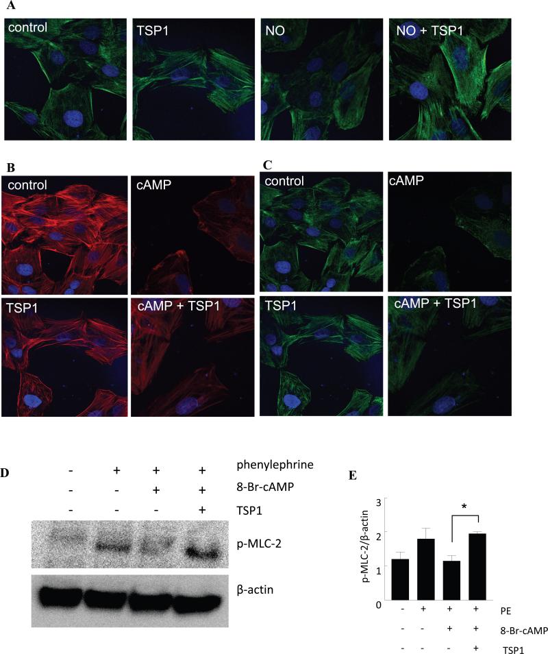
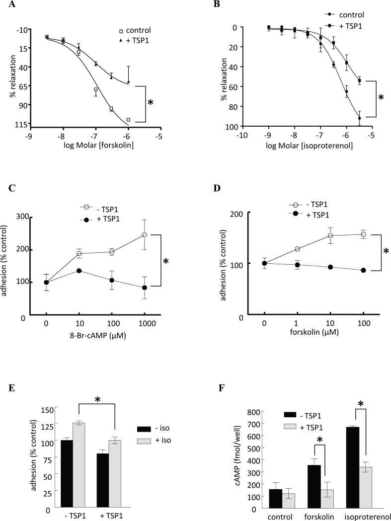
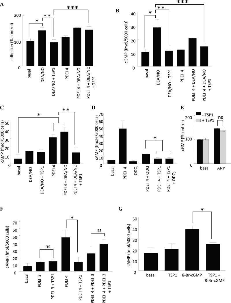
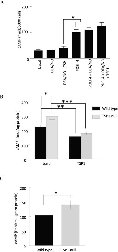
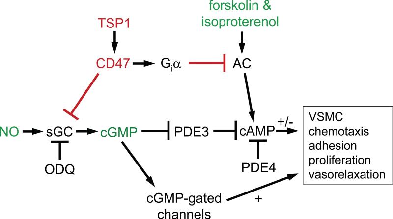
Nitric oxide (NO) drives pro-survival responses in vascular cells and limits platelet adhesion, enhancing blood flow and minimizing thrombosis. The matricellular protein thrombospondin-1 (TSP1), through interaction with its receptor CD47, inhibits soluble guanylyl cyclase (sGC) activation by NO in vascular cells. In vascular smooth muscle cells (VSMCs) both intracellular cGMP and cAMP regulate adhesion, contractility, proliferation, and migration. cGMP can regulate cAMP through feedback control of hydrolysis. Inhibition of the cAMP phosphodiesterase-4 selectively interfered with the ability of exogenous TSP1 to block NO-driven VSMC adhesion but not cGMP accumulation, suggesting that cAMP also contributes to VSMC regulation by TSP1. Inhibition of phosphodiesterase-4 was sufficient to elevate cAMP levels, and inhibiting guanylyl cyclase or phosphodiesterase-3, or adding exogenous TSP1 reversed this increase in cAMP. Thus, TSP1 regulates VSMC cAMP levels in part via cGMP-dependent inhibition of phosphodiesterase-3. Additionally basal cAMP levels were consistently elevated in both VSMCs and skeletal muscle from TSP1 null mice, and treating null cells with exogenous TSP1 suppressed cAMP levels to those of wild type cells. TSP1 inhibited both forskolin and isoproterenol stimulated increases in cAMP in VSMCs. TSP1 also abrogated forskolin and isoproterenol stimulated vasodilation. Consistent with its ability to directly limit adenylyl cyclase-activated vasodilation, TSP1 also limited cAMP-induced dephosphorylation of myosin light chain-2. These findings demonstrate that TSP1 limits both cGMP and cAMP signaling pathways and functional responses in VSMCs and arteries, by both phosphodiesterase-dependent cross talk between these second messengers and by inhibition of adenylyl cyclase activation.


Figures

Fig. 1
Fig. 1. cAMP-driven actin reorganization in VSMC is inhibited by exogenous TSP1
(A) Rat aortic VSMCs were treated in serum free medium plus 0.1% BSA (control), TSP1 (2.2 nM), DEA/NO (10μM) or DEA/NO + TSP1 (2.2 nM) for 1 h, then fixed, permeabilized and stained for p-MLC-2. Representative images three separate experiments are presented. Magnification - 60x. Cells were treated in serum free medium plus 0.1% BSA (control), TSP1 (2.2 nM), di-Bu-cAMP (100 μM) or di-Bu-cAMP + TSP1 (2.2 nM) for 1 h, then fixed, permeabilized and stained for F-actin phalloidin (B) and p-MLC-2 (C). Images are representative of three separate experiments. Magnification – 60x. (D) Aortic VSMCs at 90% confluence on 3.5 cm culture dishes were serum/additive starved over 24 h and in basal medium plus 0.1% BSA treated with TSP1 (2.2 nM) ± 8-Br-cAMP (100 μM) ± phenylephrine (10 μM), lysates prepared and Western blot analysis of p-MLC-2 determined. A representative blot of 3 separate experiments is presented. (E) Western blot expression was quantified by densitometry calculated by measuring intensity of bands using Image J and normalized to β-actin. *P<0.05, PE + 8-Br-cAMP + TSP1 versus PE + 8-Br-cAMP.
Fig. 2
Fig. 2. TSP1 inhibits cAMP-stimulated vasodilation and VSMC adhesion, and agonist-induced cAMP accumulation
Freshly harvested aortas from 12 week old male wild type C57BL/6 mice underwent disruption of the endothelium which was confirmed by lack of vasorelaxation in response to acetylcholine. Vessels were then pre-contracted and arterial vasorelaxation to a dose response curve of (A) forskolin or (B) isoproterenol ± TSP1 (2.2 nM) determined. Data represent the mean ± SD of 4 vessels in each treatment group. *P<0.005, TSP1 treated versus control. Fitted curves were analyzed by two-way ANOVA followed by the Bonferroni post test. (C) Human aortic VSMC (2 × 10⁵ cells/ml) attachment was determined on dishes coated with type I collagen (2.5 μg/ml) in the presence of the cell permeable cAMP analog 8-Br-cAMP ± TSP1 (2.2 nM). Results presented are the mean ± SD of three experiments. *P<0.05, TSP1 treated versus control. VSMC (2 x 10⁵ cells/ml) attachment was determined on dishes coated with type I collagen (2.5 μg/ml) in the presence of the indicated doses of (D) forskolin ± TSP1 (2.2 nM) or (E) isoproterenol (10 μM) ± TSP1 (2.2 nM). Results presented are the mean ± SD of three experiments. * P<0.05, TSP1 treated versus control (D); *P<0.05 TSP1 + isoproterenol versus isoproterenol (E). (F) Human aortic VSMCs were plated at 80% confluence and serum deprived over 24 h. In basal medium + 0.1% BSA cells were treated with isoproterenol (10 μM) or forskolin (1 μM) ± TSP1 (2.2 nM) and cAMP levels determined via ELISA. Results are the mean ± SD of three experiments. *P<0.05, TSP1 + isoproterenol (or forskolin) versus isoproterenol (or forskolin).
Fig. 3
Fig. 3. TSP1 regulates VSMC cAMP levels via PDE cross talk
(A) Human aortic VSMC (2 × 10⁵ cells/ml) attachment was determined on dishes coated with type I collagen (2.5 μg/ml) in the presence of the indicated treatment agents at the following concentration - DEA/NO (10 μM), PDE4 inhibitor (1 μM), TSP1 (2.2 nM). Results presented are the mean ± SD of three experiments. *P<0.05, basal versus DEA/NO; **P<0.05, DEA/NO versus DEA/NO + TSP1; ***P<0.05, DEA/NO + TSP1 versus DEA/NO + TSP1 + PDEI 4. (B) VSMCs were incubated for 5 min in basal medium (SM-GM without serum and additives) + 0.1% BSA in the presence of the indicated treatment agents at the following concentrations - DEA/NO (10 μM), PDE4 inhibitor (1 μM), TSP1 (2.2 nM) and intracellular cGMP measured by immunoassay. Results presented are representative of three experiments. *P<0.05, basal versus DEA/NO; **P<0.05, DEA/NO versus DEA/NO + TSP1; ***P<0.05, DEA/NO versus DEA/NO + TSP1 + PDEI 4. (C) VSMCs (5000 cells/well) were incubated in 96-well plates for 5 min in basal medium + 0.1% BSA in the presence of the indicated treatment agents at the following concentrations - DEA/NO (10 μM), PDE4 inhibitor (1 μM), TSP1 (2.2 nM) and intracellular cAMP measured via immunoassay . *P<0.05, basal versus PDEI 4 or DEA/NO + PDEI 4; **P<0.05, PDEI 4 or DEA/NO + PDEI 4 versus DEA/NO + TSP1 + PDEI 4. VSMCs (5000 cells/well) were incubated in 96-well plates for 5 min in basal medium + 0.1% BSA in the presence of the indicated treatment agents at the following concentrations - DEA/NO (10 μM), PDE4 inhibitor (1 μM), TSP1 (2.2 nM), ODQ (10 μM) (D) and intracellular cAMP measured via immunoassay. *P<0.05, PDEI 4 + ODQ versus PDEI 4 + TSP1 or PDEI 4 + TSP1 + ODQ. VSMCs (5000 cells/well) were incubated in 96-well plates for 5 min in basal medium + 0.1% BSA and ANP (10 μM) ± TSP1 (2.2 nM) and cGMP measured by immunoassay (E). ns = not significant, ANP versus ANP + TSP1. (F) VSMCs (5000 cells/well) were incubated in 96-well plates for 5 min in basal medium + 0.1% BSA in the presence of the indicated treatment agents at the following concentrations - DEA/NO (10 μM), PDE4 inhibitor (1 μM), PDE3 inhibitor (2 μM), TSP1 (2.2 nM) and intracellular cAMP measured via immunoassay *P<0.05, PDEI 4 versus PDEI 4 + TSP1 or PDEI 4 + PDEI 3. ns = not significant, PDEI 3 versus PDEI 3 + TSP1 and PDEI 4 + PDEI 3 versus PDEI 4 + PDEI 3 + TSP1. (G) VSMCs (5000 cells/well) were incubated in 96-well plates for 5 min in basal medium + 0.1% BSA in the presence of 8-Br-cGMP (10 μM) ± TSP1 (2.2 nM) and cAMP levels determined via immunoassay. * P<0.05, TSP1 + 8-Br-cAMP versus 8-Br-cAMP.
Fig. 4
Fig. 4. Inhibition of NO-dependent cAMP signaling by TSP1 is specific for VSMC
(A) HUVECs (5000 cells/well) were incubated in 96-well plates for 5 min in basal medium (E-GM without serum and additives) + 0.1% BSA in the presence of the indicated treatment agents at the following concentrations - DEA/NO (10 μM), PDE4 inhibitor (1 μM), TSP1 (2.2 nM) and intracellular cAMP measured via immunoassay *P<0.05, DEA/NO + TSP1 versus PDEI 4 or DEA/NO + PDEI 4. Results are representative of three separate experiments. (B) Murine derived aortic VSMCs from wild type and TSP1 null age matched C57BL/6 mice were grown to 80% surface saturation on 6-well culture plates (Nunc) and serum and growth factor deprived overnight in SM-GM (Lonza, Switzerland) and 0.1% BSA. The next morning cells were treated with TSP1 (2.2 nM) for 15 min, lysed and cAMP determined via ELISA immunoassay (GE Healthcare, Franklin, NJ). Results were normalized to total protein and are presented as the mean ± SD of three separate experiments. *P<0.05, TSP1 null versus wild type; **P<0.05, wild type + TSP1 versus wild type; ***P<0.05 TSP1 null + TSP1 versus TSP1 null. (C) Vastus lateralis skeletal muscle biopsies were obtained from age matched male C57BL/6 wild type and TSP1 null mice, snap frozen in liquid nitrogen, pulverized and suspended in lysis buffer. Following sonication cAMP levels were determined from the supernatant via ELISA immunoassay with results normalized to total protein. Results represent the mean ± SD of 6 wild type and 6 TSP1 null muscle biopsies. * P<0.05, TSP1 null versus wild type.
Fig. 5
Fig. 5. TSP1 controls VSMC response through modulating both cAMP and cGMP
TSP1 acts at the level of guanylyl cyclase (GC) to inhibit NO-induced VSMC adhesion, migration, proliferation and contraction [2,3]. NO signaling also elevates cAMP through inhibiting PDE3 activity in a cGMP-dependent manner. TSP1 can block this elevation under conditions where PDE3 is limiting for cAMP degradation. Acting through cGMP-independent pathways, TSP1 also inhibits VSMC cAMP accumulation and limits cAMP-dependent vasodilation. TSP1 control of VSMC cAMP, through cGMP cross talk and in a cGMP-independent fashion by inhibiting adenylyl cyclase activation represents additional and novel mechanisms by which TSP1 regulates VSMC contractility, arterial diameter and blood flow emphasizing the redundant nature of TSP1 regulation of vascular tone. (AC, adenylyl cyclase; Giα, G protein-coupled receptor)

References

    1. Ignarro LJ. Nitric oxide as a unique signaling molecule in the vascular system: A historical overview. J Physiol Pharmacol. 2002;53:503–514. - PubMed
    1. Isenberg JS, Wink DA, Roberts DD. Thrombospondin-1 antagonizes nitric oxide-stimulated vascular smooth muscle cell responses. Cardiovasc Res. 2006;71:785–793. - PubMed
    1. Isenberg JS, Hyodo F, Matsumoto K, Romeo MJ, Abu-Asab M, Tsokos M, Kuppusamy P, Wink DA, Krishna MC, Roberts DD. Thrombospondin-1 limits ischemic tissue survival by inhibiting nitric oxide-mediated vascular smooth muscle relaxation. Blood. 2007;109:1945–1952. - PMC - PubMed
    1. Isenberg JS, Hyodo F, Pappan LK, Abu-Asab M, Tsokos M, Krishna MC, Frazier WA, Roberts DD. Blocking thrombospondin-1/cd47 signaling alleviates deleterious effects of aging on tissue responses to ischemia. Arterioscler Thromb Vasc Biol. 2007;27:2582–2588. - PubMed
    1. Isenberg JS, Romeo MJ, Maxhimer JB, Smedley J, Frazier WA, Roberts DD. Gene silencing of cd47 and antibody ligation of thrombospondin-1 enhance ischemic tissue survival in a porcine model: Implications for human disease. Ann Surg. 2008;247:860–868. - PMC - PubMed

Publication types

MeSH terms