. 2011 Feb;32(4):1091-101.
doi: 10.1016/j.biomaterials.2010.10.003. Epub 2010 Oct 23.

Regeneration of the cavernous nerve by Sonic hedgehog using aligned peptide amphiphile nanofibers

Affiliations

Regeneration of the cavernous nerve by Sonic hedgehog using aligned peptide amphiphile nanofibers

Nicholas L Angeloni et al. Biomaterials. 2011 Feb.

Abstract

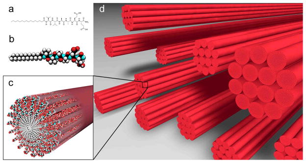
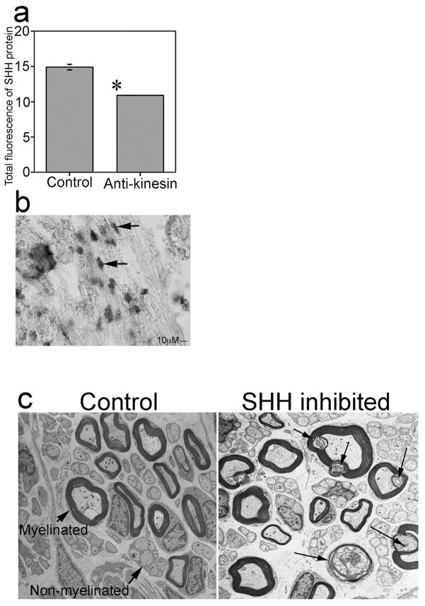
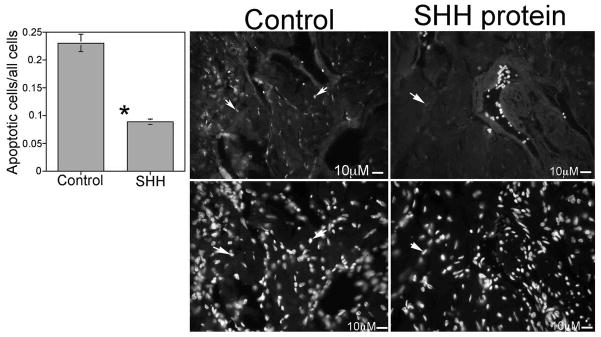
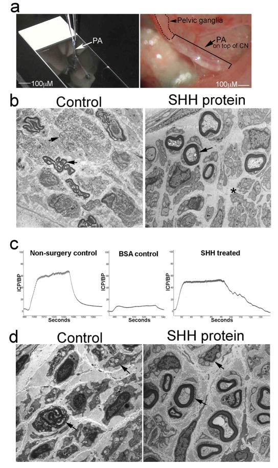
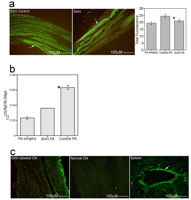
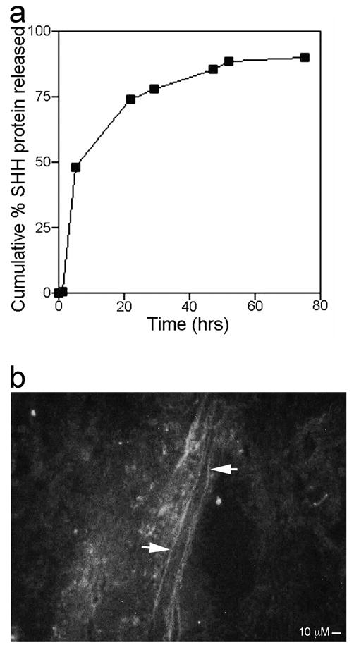
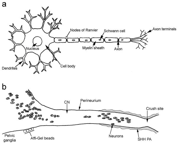
SHH plays a significant role in peripheral nerve regeneration and has clinical potential to be used as a regenerative therapy for the CN in prostatectomy patients and in other patients with neuropathy of peripheral nerves. Efforts to regenerate the cavernous nerve (CN), which provides innervation to the penis, have been minimally successful, with little translation into improved clinical outcomes. We propose that, Sonic hedgehog (SHH), is critical to maintain CN integrity, and that SHH delivered to the CN by novel peptide amphiphile (PA) nanofibers, will promote CN regeneration, restore physiological function, and prevent penile morphology changes that result in erectile dysfunction (ED). We performed localization studies, inhibition of SHH signaling in the CN, and treatment of crushed CNs with SHH protein via linear PA gels, which are an innovative extended release method of delivery. Morphological, functional and molecular analysis revealed that SHH protein is essential to maintain CN architecture, and that SHH treatment promoted CN regeneration, suppressed penile apoptosis and caused a 58% improvement in erectile function in less than half the time reported in the literature. These studies show that SHH has substantial clinical application to regenerate the CN in prostatectomy and diabetic patients, that this methodology has broad application to regenerate any peripheral nerve that SHH is necessary for maintenance of its structure, and that this nanotechnology method of protein delivery may have wide spread application as an in vivo delivery tool in many organs.


Figures

Figure 1
Figure 1
Chemical structure of the (C16)-V2A2E2-(NH2) PA used to form monodomain noodle gels (a), molecular graphics representation of the PA molecule (b), self-assembly of PA molecules into a nanofiber (c), and schematic representation of nanofiber bundles oriented in a common orientation after the noodle gel is formed (d).
Figure 2
Figure 2
(a) Quantification of SHH protein by semi-quantitative IHC analysis of penis tissue from Sprague Dawley rats that were treated with anti-kinesin or PBS in the pelvic ganglia via Affi-Gel beads. SHH protein was decreased 27% after interruption of anterograde transport in the CN by anti-kinesin. (b) In situ hybridization of Shh mRNA expression in adult Sprague Dawley rats shows Shh is localized in Schwann cells of the CN. Arrows indicate Schwann cells. 400X. (c) EM of control and SHH inhibited CN shows de-myelination and axonal degeneration of CN fibers after SHH inhibition. Arrows indicate myelinated and 15 non-myelinated fibers in control CN and myelin ovoids in SHH inhibited CN, where myelin is being broken down. 30,000X.
Figure 3
Figure 3
TUNEL assay (Top) and DAPI staining (Bottom) for all cells of penis tissue from Sprague Dawley rats that under went bilateral CN crush and were treated with either mouse IgG (control) or SHH protein by Affi-Gel beads for two weeks. SHH protein treatment of crushed CNs suppressed the apoptotic index in the penis by 53% (250X). Arrows indicate apoptotic cells.
Figure 4
Figure 4
(a) Linear PA made on a slide and placed in vivo on top of a crushed CN. 100X. (b) EM of Sprague Dawley rats that had bilateral CN crush and were treated with BSA PA (control) or SHH PA for 4 weeks. Myelinated fibers are intact in the SHH treated CN and axonal sprouts are evident in non-myelinated fibers (asterisk). Control CNs still show abundant break down of myelin in myelinated fibers and axonal degeneration in non-myelinated fibers (arrows). 30,000X and 44,000X. (c) ICP analysis of CN crushed Sprague Dawley rats that were treated with BSA PA (control) or SHH PA for 6 weeks, shows a 58% improvement in erectile function in the SHH treated rats. (d) EM of Sprague Dawley rats that had bilateral CN crush and were treated with BSA PA (control) or SHH PA for 6 weeks. The SHH PA treated rats show significant regeneration by comparison to controls as indicated by intact myelinated and non-myelinated fibers (arrows) and axonal sprouts visible in non-myelinated fibers. Control rats show the break down of myelin in myelinated fibers and degeneration of non-myelinated fibers (arrows). 70,000X and 44,000X.
Figure 5
Figure 5
(a) Quantification of GFAP protein by semi-quantitative IHC analysis in CNs from Sprague Dawley rats that under went bilateral CN crush and were treated with BSA PA (control) or SHH PA for 6 weeks, shows a 22% increase in GFAP in control BSA treated tissues that was not present in SHH PA treated CNs. GFAP increases in peripheral nerves in response to injury and decreases with regeneration. Arrows indicate GFAP protein. 160X. (b) Real time RT-PCR of Gfap RNA expression in CNs from Sprague Dawley rats that under went bilateral CN crush and were treated with BSA PA (control) and SHH PA for 6 weeks, shows a 64% increase in Gfap expression in controls and only a 36% increase in Gfap in the SHH PA treated CNs, indicating significant CN regeneration. (c) IHC assayed for CD3 (immune response marker) shows no staining present in CNs treated with SHH PA (left), or in normal CN (middle), however CD3 protein was abundant in spleen tissue (right), which was used as a positive control. 160X, 160X and 250X.
Figure 6
Figure 6
(a) Graph of cumulative percent SHH protein release from linear PA in vitro versus time (hours). Error for each point of the graph is too small for error bars to appear in the diagram. (b) Pelvic ganglia and CN from adult Sprague Dawley rats that under went bilateral CN crush and were treated with Alexa Fluor 488 labeled SHH protein via linear PA show axons between the crush site and pelvic ganglia which stain with labeled SHH protein, indicating retrograde transport of SHH protein from the crush site. 250X.
Figure 7
Figure 7
(a) Diagram of a neuron. (b) Diagram of the pelvic ganglia and cavernous nerve. CN=cavernous nerve. PA=peptide amphiphile.

References

    1. Montorsi P, Ravagnani PM, Galli S, Salonia A, Briganti A, Werba JP, et al. Association between erectile dysfunction and coronary artery disease: matching the right target with the right test in the right patient. Eur Urol. 2006;50:721–731. - PubMed
    1. User HM, Hairston JH, Zelner DJ, McKenna KE, McVary KT. Penile weight and cell subtype specific changes in a post-radical prostatectomy model of erectile dysfunction. J Urol. 2003;169:1175–1179. - PubMed
    1. Kendirci M, Hellstrom WJ. Current concepts in the management of erectile dysfunction in men with prostate cancer. Clin Prostate Cancer. 2004;3:87–92. - PubMed
    1. Perimenis P, Markou S, Gyftopoulos K, Athanasopoulos A, Giannitsas K, Barbalias G. Switching from long-term treatment with self-injections to oral sildenafil in diabetic patients with severe erectile dysfunction. Eur Urol. 2002;41:387–391. - PubMed
    1. Connolly SS, Yoo JJ, Abouheba M, Soker S, McDougal WS, Atala A. Cavernous nerve regeneration using acellular nerve grafts. World J Urol. 2008;26:333–339. - PubMed

Publication types

MeSH terms