Review
. 2011 Feb;36(2):65-77.
doi: 10.1016/j.tibs.2010.09.006. Epub 2010 Oct 23.

Protein kinases: evolution of dynamic regulatory proteins

Affiliations
Review

Protein kinases: evolution of dynamic regulatory proteins

Susan S Taylor et al. Trends Biochem Sci. 2011 Feb.

Abstract

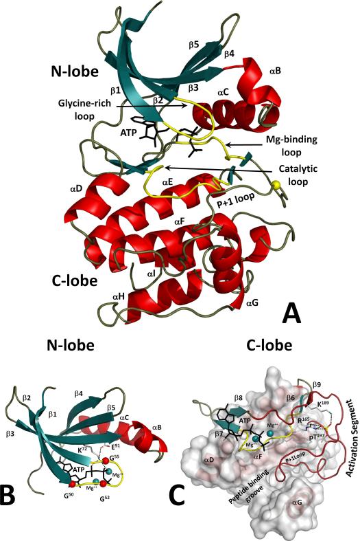
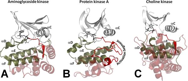
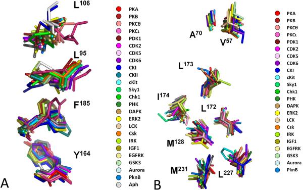
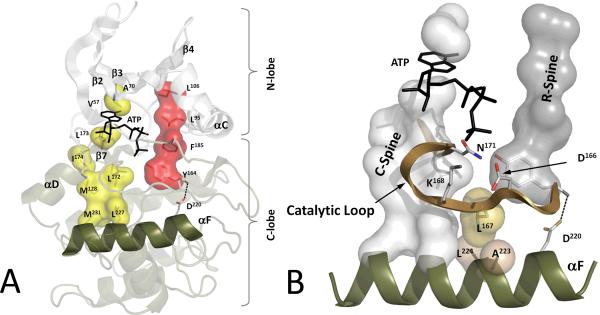
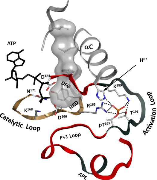
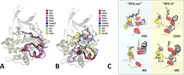
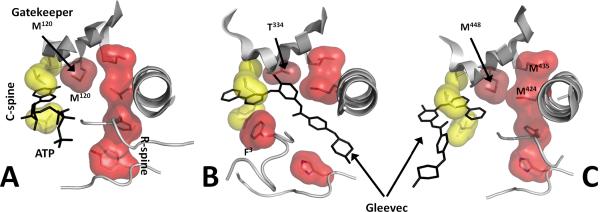
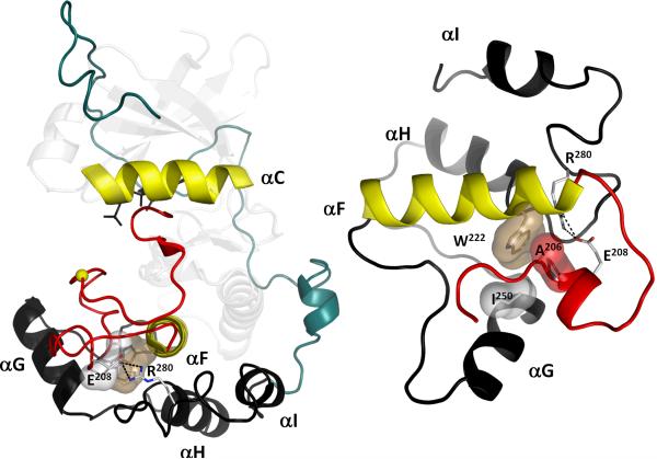
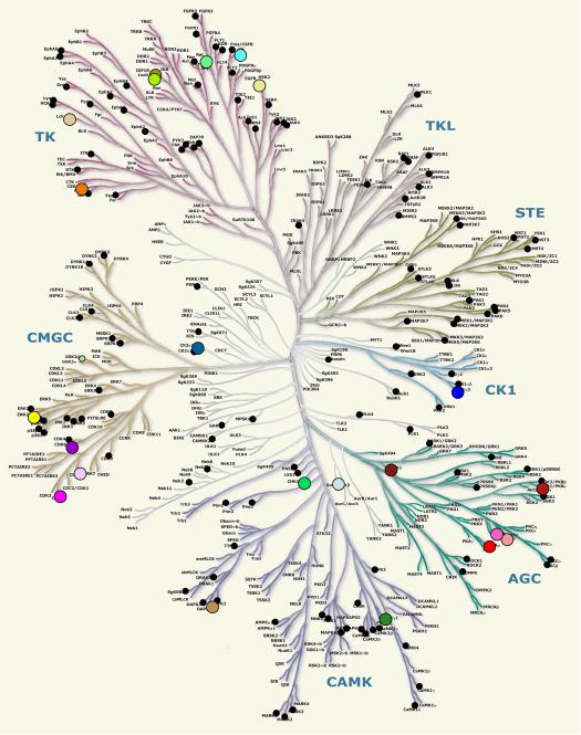
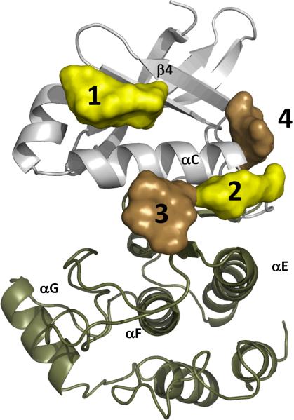
Eukayotic protein kinases evolved as a family of highly dynamic molecules with strictly organized internal architecture. A single hydrophobic F-helix serves as a central scaffold for assembly of the entire molecule. Two non-consecutive hydrophobic structures termed "spines" anchor all the elements important for catalysis to the F-helix. They make firm, but flexible, connections within the molecule, providing a high level of internal dynamics of the protein kinase. During the course of evolution, protein kinases developed a universal regulatory mechanism associated with a large activation segment that can be dynamically folded and unfolded in the course of cell functioning. Protein kinases thus represent a unique, highly dynamic, and precisely regulated set of switches that control most biological events in eukaryotic cells.


Figures

Figure 1
Figure 1. Structure of the conserved protein kinase core
A. Protein kinases have a characteristic bilobal fold. The N-terminal lobe (N-lobe) contains five β strands (1 through 5; colored teal) and a universally conserved αC-helix. The C-lobe is mostly helical (colored red). An ATP molecule is bound to a deep cleft between the lobes. Major catalytically important loops are colored yellow. B. N-lobe structure. The Glycine-rich Loop coordinates the ATP phosphates. Three conserved glycines are shown as red spheres. Lys72 from β3 couples the phosphates and the C-helix. C. Catalytic and regulatory machinery is bound to the rigid helical core of the C-lobe. The extended Activation Segment (colored dark red) contains a phosphorylation site that is bound to β9 (K189) and the HRD-arginine (R165). The P+1 Loop accommodates the P+1 residue of the peptide substrate that is docked to the peptide binding groove.
Figure 2
Figure 2. General structural differences between Eukaryote Like Kinases and EPKs
EPKs are represented by Protein kinase A (panel B). Structural elements conserved in all kinases are shown as white (N-lobe) and tan (C-lobe) cartoons. Non-conserved helical C-terminal regions of the C-lobe are shown as red transparent helices. All EPKs have a helical motif comprising three short helices: G, H and I (GHI-domain). ELKs (panels A and C) contain multiple non-conserved helices that accommodate non-peptide substrates and are specific for each kinase. Another radical difference of EPKs from ELKs is the presence of an extended Activation Segment that lies between the DFG-motif and the F-helix. This segment is the latest evolutionary feature developed by EPKs that allows dynamic regulation of their activity.
Figure 3
Figure 3. Multiple alignment of residues comprising the two Spines
All structures of active EPKs and some ELKs have conserved structural motifs in their core. They form highly conserved spatial patterns. These patterns, termed “spines”, do not form sequential motifs coming from different parts of protein kinases sequence. A. Four residues from 25 human kinases and two ELKs (PknB and Aph) form the R-spine. B. Seven residues from 24 human kinases and one ELK (PknB) form the C-spine. PKA numbering is shown. Distribution of the human kinases on the kinome tree is shown in Box 1.
Figure 4
Figure 4. The F-helix and two hydrophobic Spines define the internal architecture of the protein kinase molecule
A. The R-spine (red surface) and C-spine (yellow surface) are anchored to the F-helix (tan) in the middle of the rigid C-lobe. The C-spine is completed by the ATP molecule and, together with the R-spine, traverses the entire molecule, providing a firm, but flexible, connection between the two lobes. B. Major elements of the catalytic machinery are also anchored to the F-helix directly or via the Spines.
Figure 5
Figure 5. Phosphorylated residues in the Activation Segment control the catalytic elements of the kinase
The Activation Segment consists of a short “Magnesium binding Loop” with the DFG-motif in its N-terminus. It is followed by the most diverse part of the segment, the “Activation Loop” that contains the primary phosphorylation site (pT197). The following “P+1 Loop” forms a pocket that accommodates residues in the peptide substrate positioned immediately after the serine, threonine or tyrosine to be phosphorylated. The highly conserved Ala-Pro-Glu (APE) motif stabilizes the Activation Segment by docking to the F-helix. The primary phosphate is anchored to the immobilized HRD-arginine (R165) from the Catalytic Loop (sand ribbon). It also forms multiple bonds inside the Activation Loop and to the C-helix thereby stabilizing the active configuration of the segment that is conserved through all EPKs. Correct positioning of the DFG-phenylalanine (F185) and the C-helix assembles the Regulatory Spine (shown as a white surface), thus providing efficient catalytic activity.
Figure 6
Figure 6. Geometry of the Activation Segment in different EPK families (AGC, CMG, CAMK, STE and TK) changes significantly upon inactivation
All active EPKs have a conserved internal architecture, but inactive structures can vary significantly. (A) Activation Segments of activated kinases typically have a highly conserved conformation. (B) Different kinases can be inactivated in different ways; therefore an Activation Segment in its inactivated state can accept diverse conformations. (C) The major target of the inactivation process is the disassembly of the Regulatory Spine (red surface) and stabilization of the inactive conformation. This can be achieved in two different ways. One of the most common mechanisms is to flip the DFG motif into a “DFG out” configuration [32, 33]. One variation of this is seen in the insulin receptor and PKB where the DFG-phenylalanine (brown surface) not only flips out, but also crosses over to fill the adenine pocket site in the C-Spine (yellow surface) thereby reinforcing the inactivation by blocking ATP binding as well. In this case, the C-Helix is also flipped into an “out” conformation. There are other examples where the DFG flips “out” but does not cross over to the C-Spine. Often, however, these conformations are associated with inhibitor-bound structures so that we do not know what the conformation is in an unliganded kinase, or even in the ATP-bound inactive conformation. In other cases, the DFG appears to be in a “DFG In” state although the configuration of the DFG backbone residues is not in an active conformation (e.g. CDK2 and Src). In these cases, the R-Spine is broken by the displacement of the C-Helix residue so that the C-Helix is in an “out” conformation. Thus there are many ways to break the R-Spine.
Figure 7
Figure 7. Gatekeeper residue can stabilize the R-spine and define the affinity or binding mode of inhibitors
Gatekeeper residues are positioned at the end of the β5 strand, forming a part of the hydrophobic ATP-binding pocket and interacting with the two R-spine residues from the N-lobe. A. The gatekeeper residue (Met120) in the active form of PKA with two assembled spines (PDBID 1ATP). B. In Abl kinase the gatekeeper is a smaller threonine (Thr334) that is not an effective stabilizer of the R-spine. The latter is disassembled upon inactivation with the DFG phenylalanine flipped outside, forming an extended cavity for inhibitor (Gleevec) binding (PDBID 1OPJ). A substitution of the gatekeeper threonine to isoleucine or methionine can lock the R-spine in an active conformation [39]. C. Syk kinase not only has methionine as the gatekeeper (M448), but both residues from the R-spine that are bound to the gatekeeper are methionines (PDBID 1XBB). This, apparently, stabilizes the R-spine leading to an alternative mode of Gleevec binding.
Figure 8
Figure 8. Activation Segment and the GHI-subdomain distinguish EPKs from ELKs
An extended Activation Segment (red) is a conserved feature in EPKs. It contains several loops that perform different functions (Figure 5). This segment contains the primary phosphorylation site (yellow sphere) and is missing in ELKs (Figure 2). The GHI-subdomain (black cartoon) contains three helices G, H and I. It is also missing in ELKs, but is highly conserved in EPKs. These two structural elements surround the large hydrophobic F-helix in the middle of the C-lobe that serves as a scaffold for the protein kinase core (Figure 4). The Activation Segment and the GHI-subdomain also interact with each other via a conserved APE motif (E208-R280) which is anchored to the F-helix by hydrophobic interaction (A206, P207-W222). The AGC-specific C-terminal tail (teal cartoon) reinforces the protein kinase core stability (Box 3).
Box 1
Box 1. Structural coverage of the human kinome
The human kinome includes 518 protein kinases which were divided into seven major subfamilies [53]. The 155 publicly available human protein kinase structures were known by August 2010 (Figure I). Many of these proteins were crystallized in different conformations, bound to different ligands and proteins. A comparison of their structures demonstrates a high level of structural plasticity of protein kinases. Fig I. The human kinome. Publicly available human protein kinase structures are shown as black circles. Colored circles represent the 23 EPKs aligned in Figures 3 and 4. Kinome illustration reproduced courtesy of Cell Signaling Technology, Inc. (www.cellsignal.com).
Box 2
Box 2. Detecting functionally important residues by Local Spatial Pattern alignment
Local Spatial Pattern (LSP) alignment is a new method to compare a pair of protein molecules and to detect residues that form similar spatial patterns in both proteins. This method disregards both the sequences and main chain geometry of the proteins. All residues are considered to be individual vectors that are arrayed in space. Proteins, thus, are represented by graphs with residues as vertices and edges describing their mutual orientation. The main advantage of the method is that each residue in the detected pattern is scored according to its involvement in the formation of the pattern (Figure I). The Involvement score corresponds to the number of connections on the similarity graph. This approach allows the detection of single residues that do not form any sequential motifs, but whose spatial positions are strictly conserved. It is especially effective in recognition of spatial motifs formed by hydrophobic residues because they can easily substitute for each other and, thus, are not rigorously conserved in the protein sequence. LSP alignment played a key role in the detection of the non-consecutive hydrophobic Spines. Figure I. Involvement score is used to predict functional importance of residues. A fragment of similarity graph between PKA and CDK2 [11] showing conserved spatial relations between residues around the DFG-motif. A line (edge) between two residues (vertices) indicates that mutual spatial orientation of these residues in PKA and CDK2 are similar. Different residues can have different numbers of connections on the graph (shown in parentheses). The higher the number, the more conserved spatial relations this residue has in the molecule. This number, termed the “Involvement score”, can be used as a predictor of functional importance for the residues.
Box 3
Box 3. Stabilization of the kinase core is achieved in a kinase specific manner
Following the realization that the geometry of the kinase core is strictly defined by the F-helix and the two Spines, an important question arose: to what extent is the exterior form of the core conserved? 10 protein kinases from different families were aligned by their F-helices, and cavities on their surfaces were identified and compared [42]. Among several conserved pockets detected on the protein kinases, four surrounded the C-helix (Figure I). Pocket #1 is well known as the Hydrophobic or PIF-pocket in AGC kinases; it must be filled with a conserved hydrophobic “FxxF” motif in the C-terminus of these kinases. It also is known to be an important binding site in EGFR kinases and a docking site for cyclins in CDKs. Pocket #2 is occupied by two hydrophobic residues in the PKA N-terminus and the ERK C-terminus. Pocket #3 is another docking site for cyclin in CDKs, whereas #4 is often used in tyrosine kinases such as Src or LCK. Apparently the conserved kinase core is not stable enough and requires reinforcement by additional elements that can come from N/C-terminal tails or from other proteins. Figure I A set of conserved pockets surrounding the C-helix. The C-helix is a key structural element that, along with the Activation Segment, forms the R-spine (Figures 5, 7). Stability of this helix is vital for the kinase activity. Although the geometry and positions of these pockets can be conserved in different kinases, the way in which they are filled is a kinase-specific feature. However, the role of these interactions is the same - to stabilize the C-helix in a particular conformation.

References

    1. Krebs EG. An accidental biochemist. Annu Rev Biochem. 1998;67:xii–xxxii. - PubMed
    1. Walsh DA, Perkins JP, Krebs EG. An adenosine 3',5'-monophosphate-dependant protein kinase from rabbit skeletal muscle. J Biol Chem. 1968;243(13):3763–5. - PubMed
    1. Hunter T. Tyrosine phosphorylation: thirty years and counting. Curr Opin Cell Biol. 2009;21(2):140–6. - PMC - PubMed
    1. Barker WC, Dayhoff MO. Viral src gene products are related to the catalytic chain of mammalian cAMP-dependent protein kinase. Proc Natl Acad Sci U S A. 1982;79(9):2836–2839. - PMC - PubMed
    1. Hanks SK, Hunter T. Protein kinases 6. The eukaryotic protein kinase superfamily: kinase (catalytic) domain structure and classification. Faseb J. 1995;9(8):576–96. - PubMed

Publication types

Substances

LinkOut - more resources