. 2010 Nov 1;123(Pt 21):3796-807.
doi: 10.1242/jcs.067645.

Syndecan-1 couples the insulin-like growth factor-1 receptor to inside-out integrin activation

Affiliations

Syndecan-1 couples the insulin-like growth factor-1 receptor to inside-out integrin activation

DeannaLee M Beauvais et al. J Cell Sci. .

Abstract

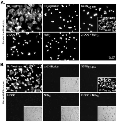
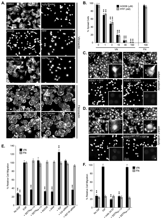
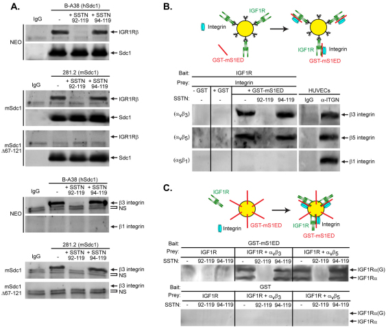
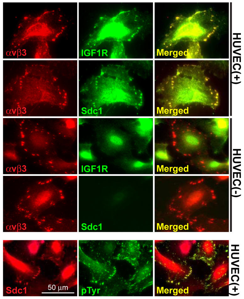
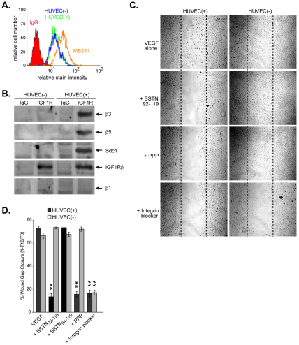
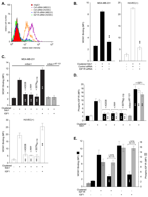
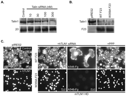
Syndecan-1 (Sdc1) engages and activates the αvβ3 (and/or αvβ5) integrin when clustered in human carcinoma and endothelial cells. Although the engagement is extracellular, the activation mechanism is cytoplasmic. This talin-dependent, inside-out signaling pathway is activated downstream of the insulin-like growth factor-1 receptor (IGF1R), whose kinase activity is triggered by Sdc1 clustering. In vitro binding assays using purified receptors suggest that association of the Sdc1 ectodomain with the integrin provides a 'docking face' for IGF1R. IGF1R docking and activation of the associated integrin is blocked by synstatin (SSTN(92-119)), a peptide derived from the integrin engagement site in Sdc1. IGF1R colocalizes with αvβ3 integrin and Sdc1 in focal contacts, but fails to associate with or activate the integrin in cells either lacking Sdc1 or expressing Sdc1(Δ67-121), a mutant that is unable to form the Sdc1-integrin-IGF1R ternary complex. Integrin activation is also blocked by IGF1R inhibitors or by silencing IGF1R or talin expression with small-interfering RNAs (siRNAs). In both cases, expression of the constitutively active talin F23 head domain rescues integrin activation. We recently reported that SSTN(92-119) blocks angiogenesis and impairs tumor growth in mice, therefore this Sdc1-mediated integrin regulatory mechanism might be a crucial regulator of disease processes known to rely on these integrins, including tumor cell metastasis and tumor-induced angiogenesis.


Figures

Fig. 1.
Fig. 1.
Sdc1-dependent activation of αvβ3 integrin is energy dependent. MDA-MB-231 cells expressing mouse Sdc1 were plated on (A) VN (20 μg/ml) or (B) antibody-coated (10 μg/ml mAb 281.2) wells in plating medium alone or in medium containing 30 μg/ml mAb LM609, 1 μM SSTN, 50 mM 2-DOG, 0.07% NaN3, or 50 mM 2-DOG plus 0.02% NaN3. Cells were incubated at 37°C for 2 hours, fixed, permeabilized and stained with Rhodamine-conjugated phalloidin or Alexa-Fluor-488-conjugated Fg, respectively. Scale bar: 50 μm.
Fig. 2.
Fig. 2.
Sdc1-dependent activation of the αvβ3 integrin requires activated IGF1R. MDA-MB-231 cells expressing mouse Sdc1 or mouse Sdc1Δ67–121 were plated on (A,B) VN or FN-coated or (C,D) Sdc1 antibody-coated wells in plating medium containing vehicle alone (DMSO), 30 μg/ml mAb LM609, 1 μM SSTN, 10 μM AG538, 10 nM PPP, 1.5 μg/ml function-blocking IGF1R mAb 24-57 or 30 ng/ml IGF1. Cells were incubated at 37°C for 2 hours, fixed, permeabilized and stained with either Rhodamine-conjugated phalloidin or Alexa-Fluor-488-conjugated Fg (insets only). Scale bar: 50 μm (mean ± s.e.m.; **P<0.01). (E,F) Non-siRNA transfected, control siRNA-transfected or IGF1R siRNA-transfected MDA-MB-231 cells were seeded on polycarbonate filters coated with either VN (black) or FN (gray) in a modified Boyden chamber. Cells were plated in plating medium alone or medium containing 1 μM SSTN, 10 μM AG538, 10 nM PPP, 1.5 μg/ml function-blocking IGF1R mAb 24-57 or 30 ng/ml IGF1. After 16 hours, cells that migrated through the filter in response to either EGF (E) or IGF1 (F) as a chemoattractant in the lower chamber were quantified by colorimetric staining (mean ± s.e.m.; **P<0.01).
Fig. 3.
Fig. 3.
Sdc1, IGF1R and the αvβ3 or αvβ5 integrin form a ternary complex that relies on the Sdc1 ectodomain. (A) Human Sdc1 and mouse Sdc1 were immunoprecipitated (using mAb B-A38 and 281.2, respectively) from MDA-MB-231 cells transfected with empty vector (NEO), mouse Sdc1 or mouse Sdc1Δ67–121. Immunoprecipitations were conducted in the presence or absence of 1 μM SSTN peptide. Blots were probed for co-precipitation of the β3 (~105 kDa), β5 (~100 kDa) or β1 (~130 kDa) integrin subunit or the IGF1Rβ (~ 95 kDa) subunit with Sdc1. Note the nonspecific bands (NS) that appear in all lanes, including the IgG isotype control precipitations. (B) IGF1R immobilized to IGF1R antibody-coated beads was incubated with purified integrin alone or purified integrin plus GST–S1ED in the presence or absence of SSTN peptide. As a comparative control, individual integrins were immunoprecipitated from HUVEC whole-cell lysates using mouse mAbs 23C6, 15F11 and HA5 (4 μg/ml) against human αvβ3, αvβ5, α5β1 integrin, respectively. Captured integrin was detected on blots by probing for the integrin β-subunits. (C) Glutathione beads bearing GST alone or GST–S1ED were incubated with purified integrin alone or purified integrin plus IGF1R in the presence or absence of SSTN peptide. Captured IGF1R was detected on blots by probing for the IGF1Rα subunit, which exists in a non-glycosylated (IGF1Rα; ~130 kDa) or glycosylated form [IGF1Rα(G); ~150 kDa].
Fig. 4.
Fig. 4.
Localization of IGF1R at the αvβ3 integrin adhesion sites requires Sdc1. Sdc1-positive and negative HUVECs were co-stained for αvβ3 integrin (mouse mAb LM609) and IGF1R (chicken anti-IGF1Rα), αvβ3 integrin (mouse mAb LM609) and Sdc1 (rabbit anti-human-S1ED) or Sdc1 (rabbit anti-human-S1ED) and anti-phosphotyrosine (mouse mAb PY20) followed by Alexa-Fluor-488 (green)- and Alexa-Fluor-546 (red)-conjugated secondary antibodies. Scale bar: 50 μm.
Fig. 5.
Fig. 5.
Sdc1 expression drives IGF1R-dependent activation of the αvβ3 integrin necessary for endothelial cell migration. (A) FACS analysis of IGF1R expression in MDA-MB-231 human mammary carcinoma cells and HUVECs against an IgG isotype control. (B) IGF1R was immunoprecipitated from HUVEC clones (grown in serum-containing medium) positive or negative for Sdc1 expression. Blots were probed for co-precipitation of the β3 (~105 kDa), β5 (~100 kDa) or β1 (~130 kDa) integrin subunit or human Sdc1 (~85 kDa). (C,D) Confluent monolayers of Sdc1-positive or negative HUVECs were serum starved before wounding and then washed twice in SFM to remove suspended cells. The cells were further cultured in SFM containing VEGF alone or VEGF plus 1 μM SSTN, 10 nM PPP or 30 μg/ml mAb LM609. The wound site was photographed immediately after wounding (T0) and again 18 hours later. Mean percentage wound closure (± s.e.m.) was calculated using the equation [1–(T18/T0)]×100. (**P<0.01).
Fig. 6.
Fig. 6.
Clustering of Sdc1 activates IGF1R leading to αvβ3 integrin activation. (A) FACS analysis for IGF1R expression in control or IGF1R siRNA-transfected MDA-MB-231 cells expressing mouse Sdc1 and HUVEC(+) cells. (B–D) Suspended cells in which mouse Sdc1 (black bars), mouse Sdc1Δ67–121 (gray bars) or human Sdc1 (open bars) or (E) IGF1R was or was not clustered (in the presence or absence of IGF1) were fixed and labeled with either WOW1 mouse Fab followed by an Alexa-Fluor-488-conjugated secondary (B,C,E) or an Alexa-Fluor-647-conjugated phospho-specific IGF1R mAb K74-218 (D,E) and analyzed by FACS. The MFI for levels of activated αvβ3 integrin (WOW1) and phosphorylated IGF1R are depicted (mean ± s.e.m.; **P<0.01).
Fig. 7.
Fig. 7.
Sdc1–IGF1R-mediated activation of the αvβ3 integrin requires talin. (A) Western blot of lysates collected from MDA-MB-231 mouse Sdc1-expressing cells transfected with control siRNA or increasing doses of human TLN1-specific siRNA and probed for expression of talin (~225 kDa; mAb TA205) or β3 integrin (~105 kDa; Fire and Ice) (B) Western blot for expression of full-length talin (mAb TA205) and HA-tagged talin head domain constructs, WT or W359A F23 (~25 kDa; anti-HA mAb 12CA5) in MDA-MB-231 mouse Sdc1-expressing cells co-transfected with human TLN1 siRNA or control siRNA (200 nM) and talin head domain or empty vector (pIRES2). (C) siRNA- and 10 nM PPP-treated cells transfected with empty vector (pIRES2) or talin head domain (WT or W359A F23) spreading on mouse Sdc1-specific mAb 281.2. Cells visualized by expression of EGFP or by staining with Alexa-Fluor-546-conjugated Fg (A546–Fg) to specifically stain activated αvβ3 integrin.

References

    1. Alexopoulou A. N., Multhaupt H. A., Couchman J. R. (2007). Syndecans in wound healing, inflammation and vascular biology. Int. J. Biochem. Cell Biol. 39, 505-528 - PubMed
    1. Anthis N. J., Wegener K. L., Ye F., Kim C., Goult B. T., Lowe E. D., Vakonakis I., Bate N., Critchley D. R., Ginsberg M. H., et al. (2009). The structure of an integrin/talin complex reveals the basis of inside-out signal transduction. EMBO J. 28, 3623-3632 - PMC - PubMed
    1. Arnaout M. A., Mahalingam B., Xiong J. P. (2005). Integrin structure, allostery, and bidirectional signaling. Annu. Rev. Cell Dev. Biol. 21, 381-410 - PubMed
    1. Askari J. A., Buckley P. A., Mould A. P., Humphries M. J. (2009). Linking integrin conformation to function. J. Cell Sci. 122, 165-170 - PMC - PubMed
    1. Avraamides C. J., Garmy-Susini B., Varner J. A. (2008). Integrins in angiogenesis and lymphangiogenesis. Nat. Rev. Cancer 8, 604-617 - PMC - PubMed

Publication types

MeSH terms