. 2010 Dec;177(6):3145-58.
doi: 10.2353/ajpath.2010.100228. Epub 2010 Oct 22.

Matriptase is involved in ErbB-2-induced prostate cancer cell invasion

Affiliations

Matriptase is involved in ErbB-2-induced prostate cancer cell invasion

Shang-Ru Wu et al. Am J Pathol. 2010 Dec.

Abstract

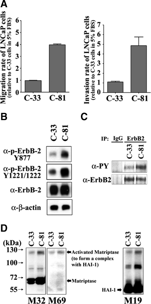
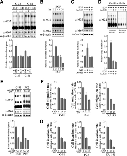
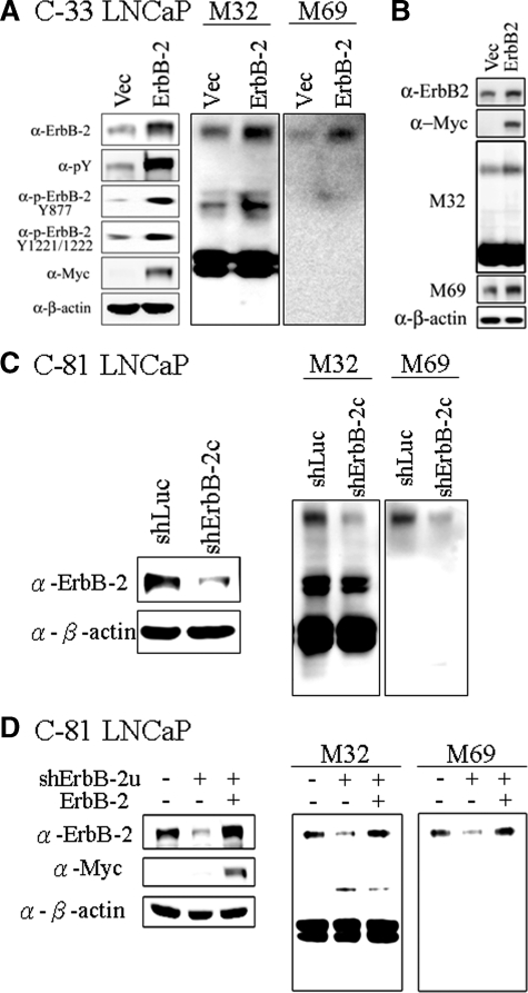
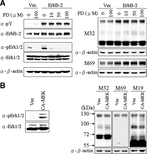
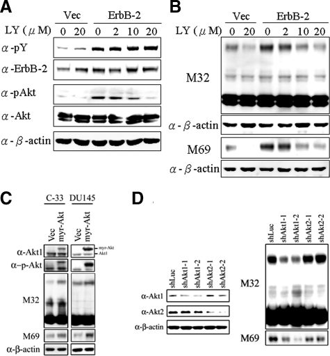
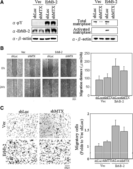
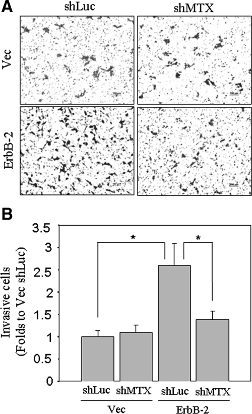
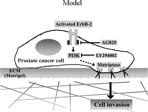
Deregulation of both ErbB-2 signaling and matriptase activity has been associated with human prostate cancer (PCa) progression. In this communication, we investigated the roles of both ErbB-2 signaling in matriptase zymogen activation and matriptase in ErbB-2-induced PCa malignancy. In a human PCa cell progression model, we observed that advanced PCa C-81 LNCaP cells exhibited an aggressive phenotype with increased cell migration and invasion capacity; these cells concurrently showed both enhanced ErbB-2 phosphorylation and increased matriptase zymogen activation compared with parental C-33 LNCaP cells. Moreover, ErbB2 activation, both ligand-dependent (eg, epidermal growth factor treatment) and ligand-independent (eg, overexpression), was able to induce matriptase zymogen activation in this cell line. Inhibition of ErbB-2 activity by either the specific inhibitor, AG825, in epidermal growth factor-treated C-33 LNCaP cells or ErbB-2 knockdown in C-81 LNCaP cells, reduced matriptase activation. These observations were confirmed by similar studies using both DU145 and PC3 cells. Together, these data suggest that ErbB-2 signaling plays an important role in matriptase zymogen activation. ErbB-2-enhanced matriptase activation was suppressed by a phosphatidylinositol 3-kinase inhibitor (ie, LY294002) but not by a MEK inhibitor (ie, PD98059). Suppression of matriptase expression by small hairpin RNA knockdown in ErbB-2-overexpressing LNCaP cells dramatically suppressed cancer cell invasion. In summary, our data indicate that ErbB-2 signaling via the phosphatidylinositol 3-kinase pathway results in up-regulated matriptase zymogen activity, which contributes to PCa cell invasion.


Figures

Figure 1
Figure 1
Migratory and invasive capabilities of different LNCaP cells and the activated levels of ErbB-2 and matriptase in those cells. A: Migratory and invasive capabilities of C-33 and C-81 LNCaP cells. After trypsinization, 4 × 10⁵ of the cells were seeded in the upper chamber of each transwell coated without or with matrigel (30 μg/cm2) in serum-free RPMI 1640 medium, and the lower chambers were filled with 5% FBS RPMI medium. Transwell migration assay was carried out for 24 hours. Migratory and invasive cells were fixed in methanol and stained with 0.25% crystal violet. Each assay was performed in triplicate for calculation of means ± SE. B: Analysis of ErbB-2 expression and phosphorylation levels by immunoblotting assays. Cell lysates were collected with 0.5% NP-40 in HEPES buffer. The ErbB-2 tyrosine phosphorylation levels were detected by using anti-phospho-ErbB-2 Tyr877 and anti-phospho-ErbB-2 Tyr-1221/Tyr-1222 antibodies (Abs). Total ErbB-2 protein level was determined by using an anti-ErbB-2 Ab (C18). C: Whole cell lysates were immunoprecipitated with an anti-ErbB-2 antibody followed by immunoblotting with anti-pTyr (4G10) and anti-ErbB-2 Abs. D: Analysis of the levels of total and activated matriptase and HAI-1 in different LNCaP cells by immunoblotting assay. LNCaP cells were lysed in 1% Triton X-100 PBS and then collected under nonboiling and nonreducing conditions. The immunoblots for total matriptase, activated matriptase, and HAI-1 were conducted by using anti-total matriptase (M32), anti-activated matriptase (M69), and anti-HAI-1 (M19) mAbs.
Figure 2
Figure 2
Effects of EGF and heregulin-β1 on matriptase zymogen activation and shedding in C-33 LNCaP cells. A: Effects of EGF and heregulin-β1 on matriptase zymogen activation in C-33 LNCaP cells. LNCaP C-33 cells were seeded at a density of 1 × 10⁶ per well in each 6-cm dish. Two days after plating, cells were starved with serum-free medium for 36 hours. After treatment with 20 ng/ml EGF or 40 ng/ml heregulin-β1 for 2 hours, cell lysates were collected under nonboiling and nonreducing conditions and immunoblotted for matriptase and activated matriptase with anti-total matriptase (M32) and anti-activated matriptase (M69) mAbs. B: Kinetics of EGF-stimulated matriptase activation in C-33 LNCaP cells. Starved cells were treated with 20 ng/ml EGF for 0, 2, 4, 8, or 24 hours. Cell lysates were then prepared and used to assay total and activated matriptase with anti-total matriptase (M32) and anti-activated matriptase (M69) mAbs. C: Serum-starved cells were treated with or without 20 ng/ml EGF in the presence or absence of 2.5 μmol/L AG825 for 2 hours. The immunoblots were performed as described in B. D: Conditioned media were collected from the cells treated with or without 20 ng/ml EGF in the presence or absence of 2.5 μmol/L AG825 for 24 hours. The immunoblots were performed as described in B. The levels of secreted matriptase were quantitated by using a densitometer and normalized to their respective cell numbers. E: The effect of AG825 on matriptase activation in C-81 LNCaP cells (left panel) and PC-3 cells (right panel). Starved cells were treated with AG825 at 0, 2.5, and 25 μmol/L for 24 hours. The immunoblots were performed as described in B. The effects of AG825 on cell migration (F) and invasion (G). After cell seeding into transwells, LNCaP C-81, PC3, and DU145 cells were treated with the indicated concentrations of AG825. Cell migration and invasion assays were performed, according to the protocol, as described in Figure 1A.
Figure 3
Figure 3
Effects of ErbB-2 level and activity on matriptase zymogen activation in PCa cells. A: Effects of ErbB-2 overexpression on matriptase activation in C-33 LNCaP cells. LNCaP C-33 cells were transiently transfected with ErbB-2 cDNA by Lipofectamine 2000 reagents, and control cells were transiently transfected with vector alone. Two days after transfection, cells were harvested for Western blot analysis. The separated proteins were transferred to a NC membrane and detected by immunoblotting with anti-pTyr (PY100), anti-ErbB-2 (C18), anti-c-Myc, and anti-β-actin (AC-15) Abs. For analyses of matriptase and HAI-1, nonreduced and nonboiled cell lysates were collected with Triton 1% X-100 in PBS, and immunoblotted with anti-total matriptase (M32) and anti-activated matriptase (M69) mAbs. B: Effects of ErbB-2 overexpression on matriptase activation in DU 145 cells. DU 145 cells were transiently transfected with ErbB-2 cDNA, and the immunoblots were performed as described in A. C: Effects of ErbB-2 knockdown on activation of matriptase in C-81 LNCaP cells. Cells were seeded at a density of 1.2 × 10⁶ per well in 6-cm dishes. One day after plating, cells were infected with lentiviral particles containing ErbB-2 shRNA for 24 hours. Control cells were infected with lentiviral particles containing luciferase shRNA. Three days after selection with 1 μg/ml puromycin, cells were harvested for Western blot analysis. For analyses of the p-Tyr and protein levels of ErbB-2, cell lysates were collected and detected by anti-pTyr (PY100) and anti-ErbB-2 (C18) Abs. For analyses of matriptase and HAI-1, nonreduced and nonboiled cell lysates were collected with Triton 1% X-100 in PBS. Matriptase and HAI-1 were detected by immunoblotting with anti-total matriptase (M32) and anti-activated matriptase (M69) mAbs. Equal protein loading was verified by blotting the membranes with an anti-β-actin Ab (AC-15). D: Effects of ErbB-2 re-expression on matriptase activation in ErbB-2-knockdown cells. C-81 LNCaP cells were infected with viral particles with control luciferase shRNA or ErbB-2u shRNA for 3 days. The ErbB-2u shRNA was designed to target a specific sequence located in the 3′ UTR of ErbB-2. The ErbB-2- knockdown cells were transfected with control vector or ErbB-2 cDNA and cultured for 3 days. Cell lysates were collected and assayed by using an anti-ErbB-2 Ab (C18). For analyses of matriptase, cell lysates were collected as described in Figure 1D to detect the levels of total and activated matriptase by immunoblotting with anti-total matriptase (M32) and anti-activated matriptase (M69) mAbs. Equal protein loading was verified by blotting the membranes with an anti-β-actin Ab (AC-15).
Figure 4
Figure 4
Role of MEK/ERKs in ErbB-2-induced matriptase activation in C-33 LNCaP cells. A: Stable ErbB-2-overexpressing C-33 LNCaP cells were seeded at a density of 6 × 10⁵ per well in a 6-well plate. Two days after plating, cells were treated with PD98059 at concentrations of 0, 10, 50, and 100 μmol/L; control transfected cells were treated with concentrations of 0 and 100 μmol/L PD98059. Treatment was carried out for 24 hours, and then cells were harvested for Western blot analysis. Cell lysates were collected with 0.5% NP-40 in HEPES buffer. The p-Tyr and protein levels of ErbB-2 were analyzed by immunoblotting with anti-pTyr (PY100) and anti-ErbB-2 (C18) Abs. The phosphorylation and protein levels of Erk1/2 were detected by anti-phosphoErk (Thr202 and Tyr204 of Erk1; Thr185 and Tyr187 of Erk2) and anti-Erk1/2 Abs. Nonreduced and nonboiled cell lysates were used for immunoblots to detect the levels of total and activated matriptase with anti-total matriptase (M32) and anti-activated matriptase (M69) mAbs. Equal loading was evaluated with an anti-β-actin Ab. B: Effects of a constitutively active MEK on matriptase activation in C-33 LNCaP cells. Cells were seeded at a density of 1.2 × 10⁶ per well in 6-cm dishes. Two days after plating, cells were transiently transfected with a constitutively active MEK (CA-MEK) cDNA and harvested 48 hours after transfection; control cells were transfected with vector alone. Cell lysates were collected with 0.5% NP-40 in HEPES buffer. The phosphorylation and protein levels of Erk1/2 were analyzed by immunoblotting with an anti-phosphoErk (Thr202 and Tyr204 of Erk1; Thr185 and Tyr187 of Erk2) and anti-Erk1/2 Abs. Nonreduced and nonboiled cell lysates were used to assay the activation status and total matriptase levels with anti-total matriptase (M32) and anti-activated matriptase (M69) mAbs. An anti-β-actin (AC-15) Ab was used to evaluate protein loading.
Figure 5
Figure 5
Role of PI 3 kinases/Akt in ErbB-2-induced matriptase activation in C-33 LNCaP cells. Stable ErbB-2-overexpressing C-33 LNCaP cells were seeded at a density of 6 × 10⁵ per well in a 6-well plate. Two days after plating, cells were treated with LY294002 at concentrations of 0, 2, 10, and 20 μmol/L; control cells were treated with concentrations of 0 and 20 μmol/L PD98059. Treatment was carried out for 24 hours, and then cells were harvested for Western blot analysis. A: Cell lysates were collected with 0.5% NP-40 in HEPES buffer and evaluated by immunoblots for p-Tyr and protein levels of ErbB-2, performed as described previously. The phosphorylation and protein levels of Akt were detected by anti-phosphoAkt (Ser473) and anti-Akt1/2 Abs. B: Nonreduced and nonboiled cell lysates were used for immunoblotting to detect the activation status and total matriptase with anti-total matriptase (M32) and anti-activated matriptase (M69) mAbs. An anti-β-actin (AC-15) Ab was used to evaluate protein loading. C: Role of a constitutively activated Akt (Myr-Akt) in matriptase zymogen activation in C-33 LNCaP cells and DU 145 cells. LNCaP C-33 cells and DU 145 cells were transiently transfected with Myr-Akt and control plasmids by using Lipofectamine 2000. Two days after transfection, cell lysates were collected and analyzed by immunoblotting with anti-phosphoAkt (Ser473), anti-Akt1/2, anti-total matriptase (M32), and anti-activated matriptase (M69) Abs, respectively. D: Effect of Akt1 or Akt2 knockdown on activation of matriptase in LNCaP C-81 cells. Cells were seeded at a density of 1.2 × 10⁶ per well in 6-cm dishes. One day after plating, cells were transfect with Akt1 shRNAs (shAkt1-1 and shAkt1-2), Akt2 shRNAs (shAkt2-1 and shAkt2-2), and control luciferase shRNA with Lipofectamine 2000. Cell lysates were collected for analysis of the levels of Akt1 and Akt2 protein by immunoblot with anti-Akt1 and anti-Akt2 Abs. For analyses of matriptase, cell lysates were collected and immunoblotted with anti-total matriptase (M32) and anti-activated matriptase (M69) mAbs. Equal protein loading was assessed with an anti-β-actin Ab (AC-15).
Figure 6
Figure 6
Effects of matriptase knockdown on ErbB-2-promoted cell migration of prostate cancer cells. Cells were seeded at a density of 1.2 × 10⁶ per well in 6-cm dishes. Cells were infected by lentiviral particles with shRNAs specific to matriptase for 24 hours, selected by 1 μg/ml puromycin for 72 hours and then harvested for Western blot analysis. A: Cell lysates were collected with 0.5% NP-40 in HEPES buffer. The p-Tyr and protein levels of ErbB-2 were detected by anti-pTyr (PY100) and anti-ErbB-2 (C18) Abs. Nonreduced and nonboiled cell lysates were used for immunoblotting to detect the activation status and total matriptase with anti-total matriptase (M32) and anti-activated matriptase (M69) mAbs. Loading was analyzed with an anti-β-actin mAb (AC-15). B: Effects of matriptase knockdown on ErbB-2-promoting cell motility by wound-healing assays. Wounds with widths of approximately 250 μm were made by scraping by using 10-μl pipette tips. Cells were incubated for 24 hours for wound-healing assay. Images were captured by a light microscopy with a magnification of 100×. The dotted lines define the edges of the wounds. Migratory distances (widths at 0 hours to widths at 24 hours) were analyzed by a NIS-Elements D software (Nikon) and are represented as means ± SE calculated from triplicates; a statistically significant difference (*P < 0.05) was observed between Vec/shLuc and ErbB-2/shLuc. C: Effects of matriptase knockdown on ErbB-2-promoting cell motility by transmigration assays. After trypsinization, 1 × 10⁵ cells were seeded with serum-free RPMI 1640 medium in each of the upper chambers, and the lower chambers were filled with 10% FBS RPMI 1640 medium. Transwell migration assay was carried out for 48 hours. Migratory cells were fixed in methanol and stained with 1% crystal violet, and images were captured by a light microscopy (original magnification, ×100). Amounts of migratory cells on each filter were counted from eight random fields (original magnification, ×200). Each assay was performed in triplicate for calculation of means ± SE; a statistically significant difference, *P < 0.05 was observed between Vec shLuc and ErbB-2 shLuc.
Figure 7
Figure 7
Invasion assay of ErbB-2-overexpressing LNCaP cells with or without matriptase knockdown. Matriptase knockdown was performed as described in Figure 6A. For cell invasion assays, each filter insert was coated with 30 μg/cm2 matrigel. After trypsinization, 1 × 10⁵ cells were seeded with serum-free RPMI 1640 medium in each insert chamber, and lower chambers were filled with 10% FBS RPMI 1640 medium. A: Transwell invasion assays were carried out for 48 hours. Invasive cells were fixed in methanol and stained with 1% crystal violet. Images were captured by a light microscopy (original magnification, ×100). B: Numbers of invasive cells on each filter were counted from eight random fields (original magnification, ×200). Each assay was performed in triplicate for calculation of means ± SE; statistically significant differences, *P < 0.05 were observed between Vec/shLuc and ErbB-2/shLuc, as well as ErbB-2/shLuc and ErbB-2/shMTX.
Figure 8
Figure 8
A model for the role of matriptase in ErbB-2-driven prostate cancer cell invasion. ErbB-2 hyperactivation by EGF stimulation or receptor overexpression results in the up-regulation of matriptase zymogen activation, at least in part via PI 3 kinase, leading to enhanced prostate cancer cell invasion.

Similar articles

Cited by

References

    1. Chen CJ, You SL, Lin LH, Hsu WL, Yang YW. Cancer epidemiology and control in Taiwan: a brief review. Jpn J Clin Oncol. 2002;32 Suppl:S66–S81. - PubMed
    1. Jemal A, Siegel R, Ward E, Murray T, Xu J, Thun MJ. Cancer statistics, 2007. CA Cancer J Clin. 2007;57:43–66. - PubMed
    1. Sporn MB. The war on cancer. Lancet. 1996;347:1377–1381. - PubMed
    1. Stetler-Stevenson WG, Aznavoorian S, Liotta LA. Tumor cell interactions with the extracellular matrix during invasion and metastasis. Annu Rev Cell Biol. 1993;9:541–573. - PubMed
    1. Carles J, Lloreta J, Salido M, Font A, Suarez M, Baena V, Nogue M, Domenech M, Fabregat X. Her-2/neu expression in prostate cancer: a dynamic process? Clin Cancer Res. 2004;10:4742–4745. - PubMed

Publication types

MeSH terms